1 min read 0

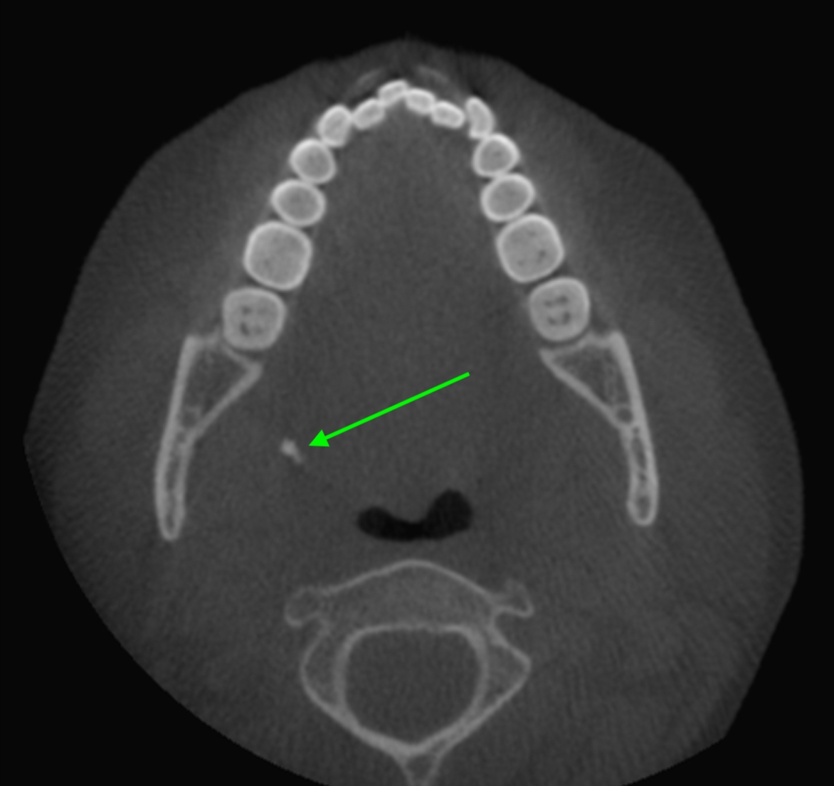
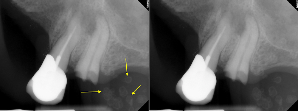
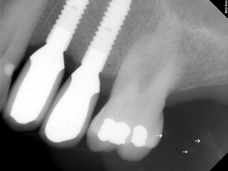
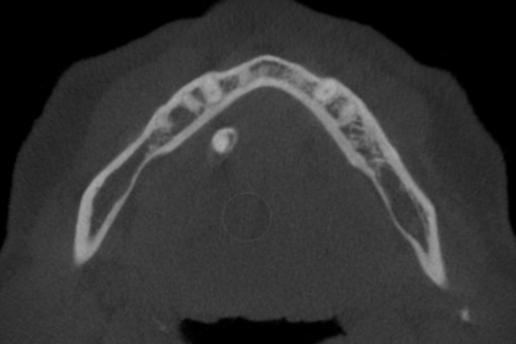
Case of the Week: Sialolith with layers (CBCT)

This week I have a case of a sialolith on CBCT (still images and videos) showing the layers of calcifications as it grows. First some information on sialoliths. QUICK DEFINITION: Sialoliths are calcifications within the ducts of the salivary glands. Most commonly associated with the submandibular salivary gland in the…
1 min read 0

Case of the Week: Tonsiliths on CBCT

Being that I realized I need to update (and add) the interpretation pages as many of them don’t have CBCT examples or information. I thought I’d start with something I come across nearly every week – tonsiliths. This week I have two cases of tonsiliths seen on CBCT. Tonsiliths are…
1 min read 1

Case of the Week: Osteoma Cutis

Happy New Year! This is a fun case that would always stump the students on the exact name of this entity.  We also came across it so often that one student crossed out the description of rare in their radiology book to use in the viewing room. 🙂 Osteoma cutis…
1 min read 0

Case of the Week: Osteoma Cutis (2D & CBCT)

This week I have a fun case of osteoma cutis on both 2D images and CBCT images. Osteoma cutis is an ossification within the dermis. It presents on 2D radiographs as punctate, round/ovoid, radiopaque entities.  Most of the cases I come across it occurs in multiples.  First is the 2D radiographs…
1 min read 0

Case of the Week: Sialolith

This week I have a neat example of a sialolith on a pantomograph.  This was an incidental finding at a new patient exam.  Sialoliths tend to be single, but this case has multiple calcifications within the duct of the submandibular salivary gland.  On the pantomograph there are multiple, irregular shaped…