5 min read 0

Anatomy Monday: Anatomy on Mandibular Periapical Radiographs

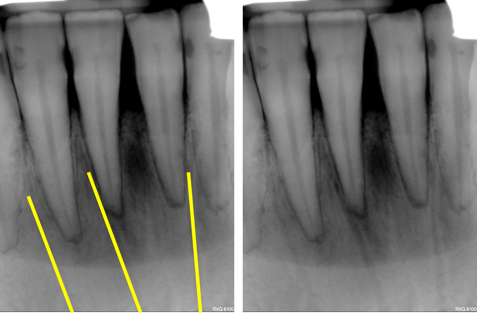
It’s been a while since I’ve updated anatomy seen on intraoral radiographs so I’m continuing with all the anatomy visible on mandibular periapical radiographs. Some of these were seen in the recent posts on canals and foramina but this will be the one stop spot for everything mandibular periapical radiographs.…
3 min read 1

Anatomy Monday: Canals on Intraoral Radiographs

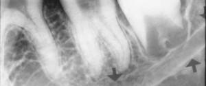
Since a couple of weeks ago I went over foramina on intraoral radiographs (periapical radiographs and bitewings), I thought I’d keep on going with more and this time go over canals. There is 4 again with 2 in the maxilla and 2 in the mandible; however one of the two…
1 min read 0

Anatomy Monday: Mandibular Canal

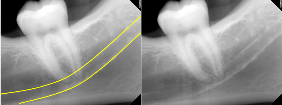
This week and month is going to be canals and ducts visible on intraoral and extraoral radiographs. I’m starting with a canal frequently sought out specifically for implants and extractions; the mandibular canal.  The mandibular canal is visible on both intraoral radiographs and extraoral radiographs. It presents as a radiolucent…
3 min read 5

Anatomy on Radiographs: Pantomographs Part 1

Ok, I am back and now onto pantomographs. Since these images are larger and will take up much more space I have decided to break them down into 2 parts.  I also will have several anatomy identified on each radiograph as well.  Here we go. Mandible *The mandibular condyle (two…
2 min read 7

Anatomy on Radiographs: Intraoral Radiographs Part 2

This is part 2 (posterior) of anatomy on intraoral radiographs. Mandible The mental foramen  appears as a round to oval radiolucent area near the apex of the second premolar. The inferior alveolar nerve canal (mandibular canal) appears as radiolucent band with two thin radiopaque lines running parallel to each other…