1 min read 0

Anatomy Monday: Mandibular Canal

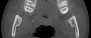
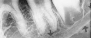
This week and month is going to be canals and ducts visible on intraoral and extraoral radiographs. I’m starting with a canal frequently sought out specifically for implants and extractions; the mandibular canal.  The mandibular canal is visible on both intraoral radiographs and extraoral radiographs. It presents as a radiolucent…
1 min read 3

Anatomy Monday: Nasolabial Fold (soft tissue)

This week is another soft tissue entity that can be seen on both intraoral and extraoral radiographs. The nasolabial fold presents as a diagonal transition line. A transition line is seen as a defined line where part of the radiograph appears more radiopaque due to superimposition of soft tissue. The…
1 min read 7

Anatomy Monday: Anterior Nasal Spine (maxilla)

This week’s anatomy is the anterior nasal spine which is found in the midline at the floor of the nasal cavity as a bony protuberance where the right and left maxilla meet (as seen on the skull below – orange arrow).On radiographs, this will appear as an inverted triangle or…
1 min read 0

Anatomy Monday: Coronoid Process (mandible)

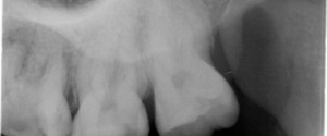
The coronoid process is the superior aspect of the anterior portion of the ramus of the mandible.  It is visible on intraoral radiographs but not mandibular periapical radiographs instead it is seen on maxillary molar periapical radiographs.  This typically due to the posterior placement of the image receptor and holder…
1 min read 0

Anatomy Monday: Vascular/Nutrient Canals (mandible)

This Monday I am showing vascular canals or nutrient canals as they are sometimes referred to. While they are in both the maxilla and mandible, they are more commonly seen in the mandible particularly the anterior. This is due to thin bone in this area. They will appear as radiolucent…