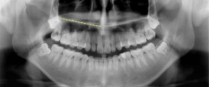
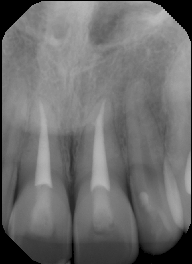
1 min read 1

Anatomy Monday: Floor of the nasal cavity (maxilla)

The floor of the nasal cavity that is visible on radiographs is actually the junction of the floor and lateral wall or vomer.  On intraoral radiographs this will appear as a single radiopaque straight line superior to the maxillary teeth.  It is visible on any periapical radiograph (anterior and posterior). …
1 min read 6

Anatomy Monday: Y line of Ennis (maxilla)

Today I am starting a new series for Mondays on anatomy. I will be showing different anatomical landmarks for both intraoral and extraoral radiographs.  This first entity I am showing is a radiographic anatomical landmark; the Y line of Ennis.  This is sometimes referred to as an Inverted Y.  It…
3 min read 5

Anatomy on Radiographs: Pantomographs Part 1

Ok, I am back and now onto pantomographs. Since these images are larger and will take up much more space I have decided to break them down into 2 parts.  I also will have several anatomy identified on each radiograph as well.  Here we go. Mandible *The mandibular condyle (two…