1 min read 6

Case of the week: Talon cusp with clinical photo

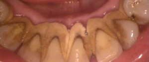
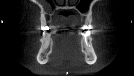
This week I have a case of a large talon cusp on a maxillary central incisor along with a clinical photo.  A talon cusp is a hyperplasia of the cingulum of a tooth and typically classified as a variant of normal anatomy.  In the anterior, it will present as a…
1 min read 0

Case of the week: Calculus with clinical photo

This week I have a case of some serious calculus on the mandibular anterior teeth.  I have both a radiograph and a clinical photo as well.  Calculus appears a radiopaque ‘growths’ on the mesial and/or distal surfaces of teeth.  It is typically found near the cemento-enamel junction but can occur…
1 min read 0

Case of the Week: Dens invaginatus with clinical photo

This week I have a case of dens invaginatus.  I have shown a case of dens invaginatus before but this time I have a clinical photo to show the infolding of the enamel.  This case is involving a mandibular premolar.  The radiograph shows a radiopaque inverted triangle with a radiolucent…
1 min read 0

Case of the Week: Enamel pearl

This week I am showing a case of an enamel pearl along with the clinical photo of the tooth after it was extracted.  Enamel pearls are an extra ‘blob’ of enamel near the furcation area on molars.   They will present as circular radiopaque areas near the furcation.   They…