1 min read 4

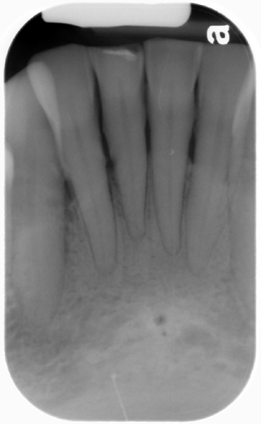
Case of the week: Double Lingual Foramina

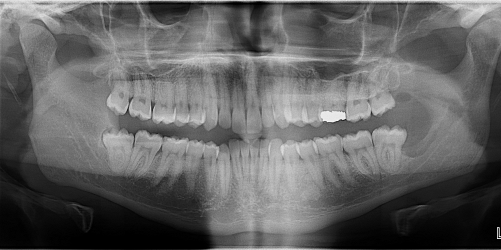
This weeks case is an interesting variant of normal anatomy – two lingual foramina seen on a periapical radiograph.  Some patients can have up to three lingual foramina. Note the two well-defined, circular radiolucent entities inferior to the mandibular central incisors.  The superior one is larger than the inferior one.…
2 min read 0

Anatomy on Radiographs: Occlusal Radiographs

This is the last post I have planned for anatomy and it will be on occlusal radiographs. Mandible Short and sweet here with only one anatomical landmark readily visible. The genial tubercles (yellow arrow) appear as a small radiopaque mass(es) extending off the lingual aspect of the mandible. Maxilla The nasal…
2 min read 7

Anatomy on Radiographs: Intraoral Radiographs Part 2

This is part 2 (posterior) of anatomy on intraoral radiographs. Mandible The mental foramen  appears as a round to oval radiolucent area near the apex of the second premolar. The inferior alveolar nerve canal (mandibular canal) appears as radiolucent band with two thin radiopaque lines running parallel to each other…
1 min read 0

Case of the Week: Blunderbuss apex

This week I wanted to show normal anatomy of a developing tooth; the blunderbuss apex.  A blunderbuss apex is an open apex of a developing tooth. It is named blunderbuss due to the similar appearance to a blunderbuss rifle.  An open or blunderbuss apex appears as pyramidal radiolucent area.  This…