1 min read 24

Caries Interpretation: Interproximal Caries

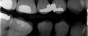
Today is the start of a series of posts on caries interpretation which can be challenging at times. Today is interproximal caries. Here’s the goods. Most commonly found at the level of the contact point or slightly apical to it. Frequently occurs as a radiolucent triangle in the enamel with…
1 min read 2

Anatomy Monday: Gubernaculum Dentis

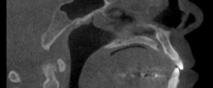
This week I have a case of anatomy that I more commonly see on CBCT images but have occasionally come across on 2D radiographs; the gubernaculum dentis. The gubernaculum dentis is a canal of connective tissue from a developing tooth bud to the overlying mucosa. It presents as a radiolucent…
1 min read 0

Anatomy Monday: Paradoxic Curvature

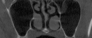
This week is the last anatomy I’ll be showing from my book (Interpretation Basics of Cone Beam Computed Tomography).  It’s another aspect of the nasal cavity involving the middle concha. Paradoxic curvature is when the curve of the concha is towards the nasal septum instead of the lateral border of…
1 min read 2

Anatomy Monday: Pineal Gland Calcification

This week I’m onto a calcification encountered in the soft tissue of the brain; the pineal gland.  It appears as a well-defined radiopaque entity in the mid-line of the brain superior to the spinal column. Another entity from my book (Interpretation Basics of Cone Beam Computed Tomography). If you have…
1 min read 5

Case of the Week: Pulp Stones

This is an interesting case of multiple teeth with pulp stones. Pulp stones present as well-defined radiopaque entities in the pulp chamber and/or root canal space.  If they are large enough, the canal will be enlarged around them to accommodate them.  Check out the pulp stone party below. 😀 If…