1 min read
6
Anatomy Monday: Hamulus
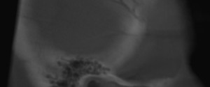
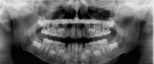
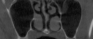
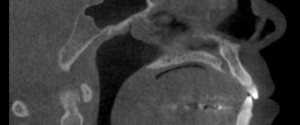
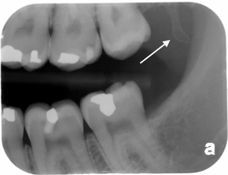
The hamulus is always a fun find when I’m working with the students on interpretations as it really forces them to recall anatomy beyond just the maxilla and mandible. The hamulus is a small bony hook that projects off the medial pterygoid plate of the sphenoid bone (that’s a tongue…