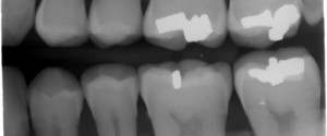
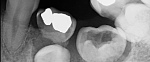
1 min read 3

Case of the Week: Microdont

This week is a fun case of a microdont third molar. On the bitewing radiograph it’s difficult to see that it’s a smaller than normal sized tooth but the periapical radiograph shows it nicely. If you have any questions or comments on microdonts, please leave a comment or question below.…
1 min read 0

Lateral Cephalometric Skull Anatomy – Part VI

This is the final post and it will cover soft tissue anatomy seen on lateral cephalometric skull radiographs. Glabella (yellow dotted arrow) Nasion (white solid arrow) Pronasale (yellow solid arrow) Subnasale (black dotted arrow) Labrale superius (turquoise dotted arrow) Stomion (green solid arrow) Labrale inferius (turquoise solid arrow) Gnathion (white…
1 min read 0

Lateral Cephalometric Skull Anatomy – Part V

Today is all about the mandible. Condyle (black curved dotted line) Coronoid process (white triangular dotted line) Mandibular first molar (green oval) Infradentale (yellow dotted arrow) – Superior facial bone height near the level of the cemento-enamel junction of the mandibular incisors. B point (white solid arrow) – The posteriormost…
1 min read 3

Anatomy Monday: Nasolabial Fold (soft tissue)

This week is another soft tissue entity that can be seen on both intraoral and extraoral radiographs. The nasolabial fold presents as a diagonal transition line. A transition line is seen as a defined line where part of the radiograph appears more radiopaque due to superimposition of soft tissue. The…
1 min read 5

Case of the Week: Ectopic Eruption & Internal Resorption

This week I have a fun case of 2 finds on 1 radiograph. This bitewing radiograph shows internal resorption (enlarged pulp chamber) of the maxillary left deciduous second molar and ectopic eruption of the mandibular left first premolar into the deciduous second molar.  Check it out below. 🙂 If you…