This week I have a case of sinusitis on cone beam CT along with another educational video. 🙂
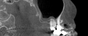
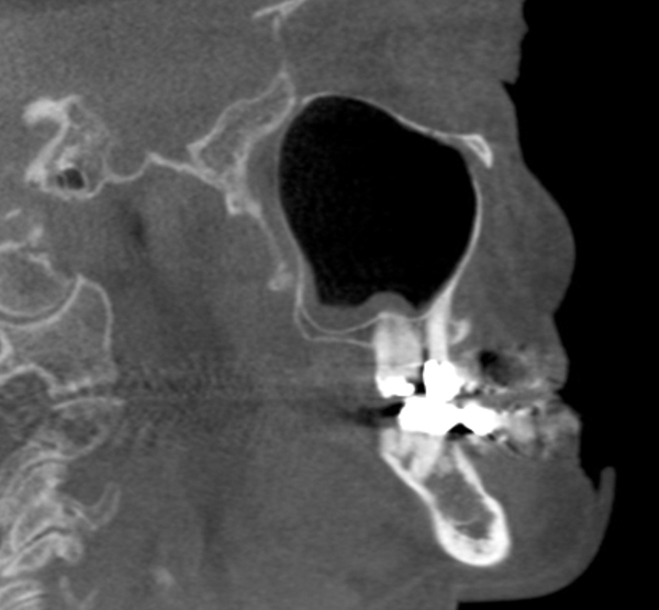
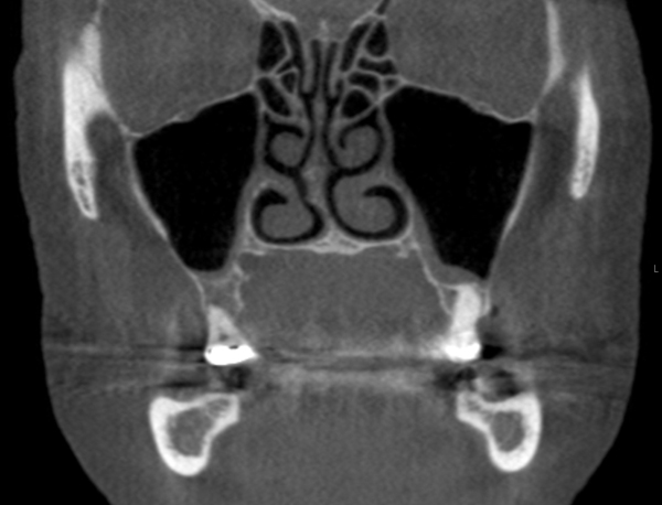
On the cone beam CT images note the radiopaque band that follows the floor and the posterior border on the sagittal view and the floor and medial border of the left maxillary sinus on the coronal view. There is also a slight thickening of the right maxillary sinus evident on the coronal view.
And now onto another fun educational video about sinusitis by Dental Class of 2015 students Derek Hoffman and Cory Wilkinson.
If you have any questions about sinusitis or the case shown above, please leave them in the comments below. Thanks and enjoy!


