I’m updating Anatomy Monday to Anatomy is your friend (I didn’t want to feel tied to only posting on Monday – but go figure this initial one is coming out on a Monday :P).
I thought I’d start by picking a random periapical radiograph and identifying all the anatomy visible on this image (there may be more anatomy visible on other similar images of this area but this single image has what it has). Below is a list of the anatomy covered in the post if you want to jump to any specific one.
- intermaxillary suture
- soft tissue tip of the nose
- anterior nasal spine
- floor of the nasal cavity
- superior foramina of the nasopalatine canal
- nasal septum
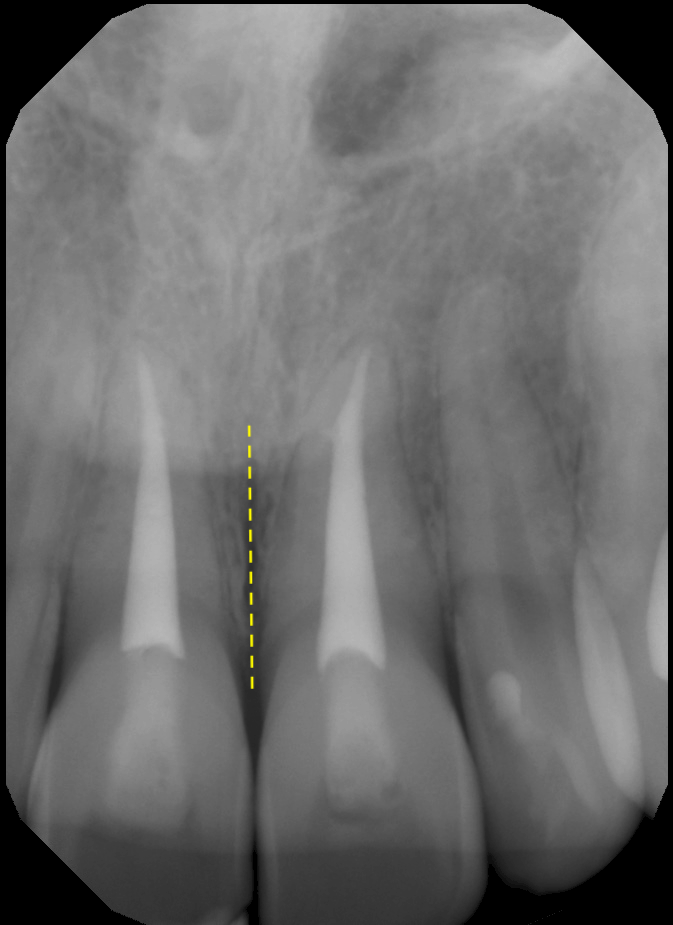
Intermaxillary Suture
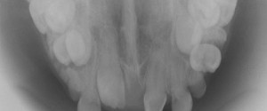
The intermaxillary suture is between the maxillary central incisors. It has varying levels of fusion. In adults, it may not be evident if it is completely fused. It appears as a vertical radiolucent line between the maxillary central incisors on the midline.
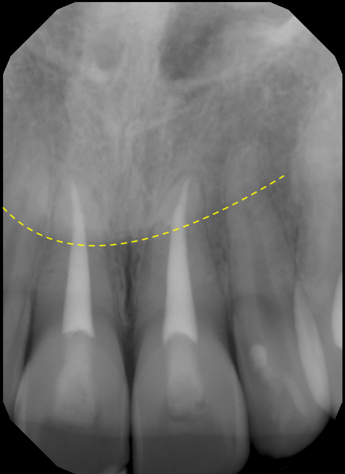
Tip of the Nose (soft tissue)
This will appear as a transition line on a 2D radiograph. It has a curved appearance over the roots of the maxillary central incisors.
Anterior Nasal Spine
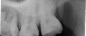
The anterior nasal spine is a triangular projection in the midline at the level of the floor of the nasal cavity. Due to the inferior angulation of the x rays, this will appear as an inverted triangle or V-shaped radiopaque area in the midline superior to the maxillary central incisors.
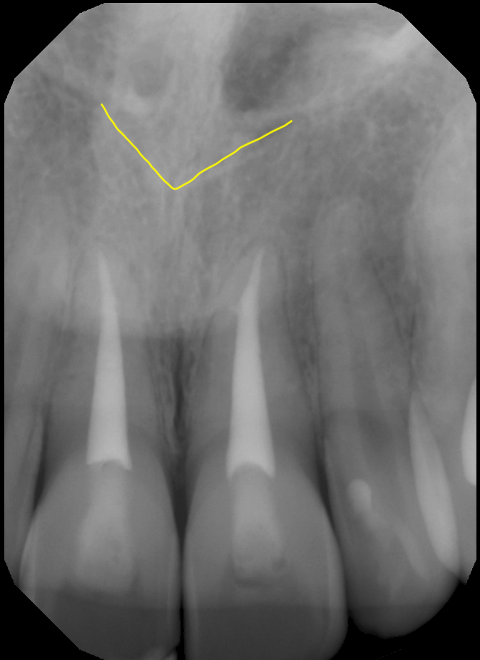
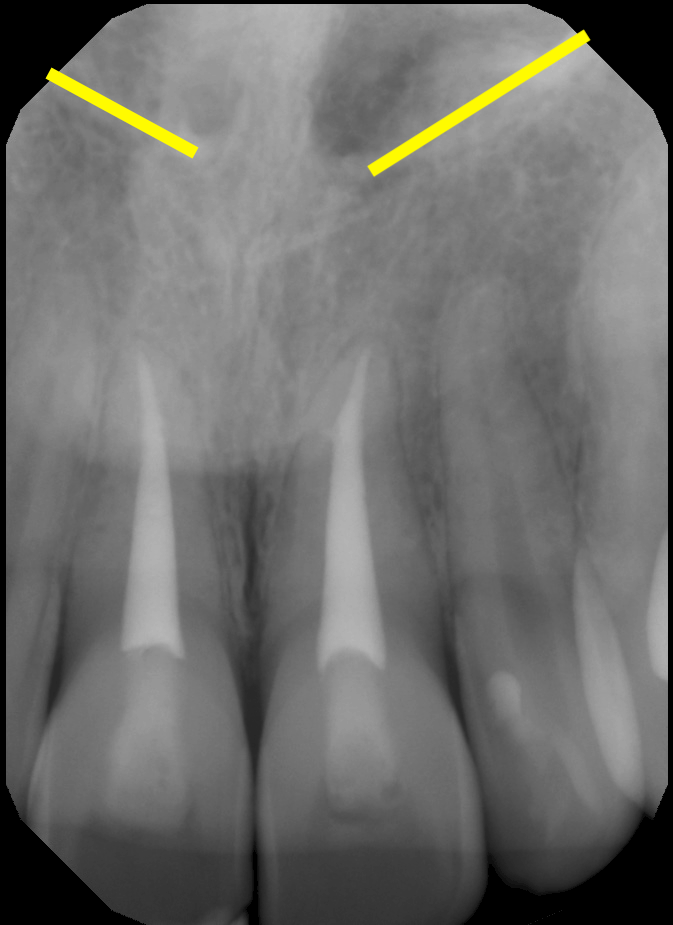
Floor of the Nasal Cavity
The floor of the nasal cavity presents as oblique radiopaque lines/bands coming off the anterior nasal spine laterally.
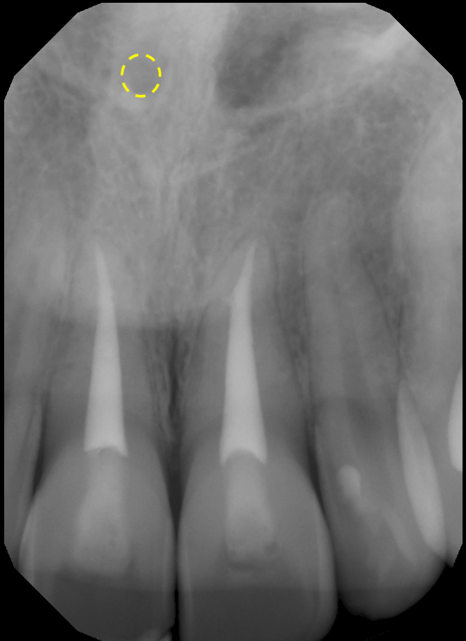
Superior Foramina of the Nasopalatine Canal
The superior foramina of the nasopalatine canal are on of the less frequent anatomy I come across on 2D radiographs. There are 2, one each just lateral to the nasal septum. They appear as a circular/ovoid radiolucent area. Sometimes only one of them is visible – as in this case.
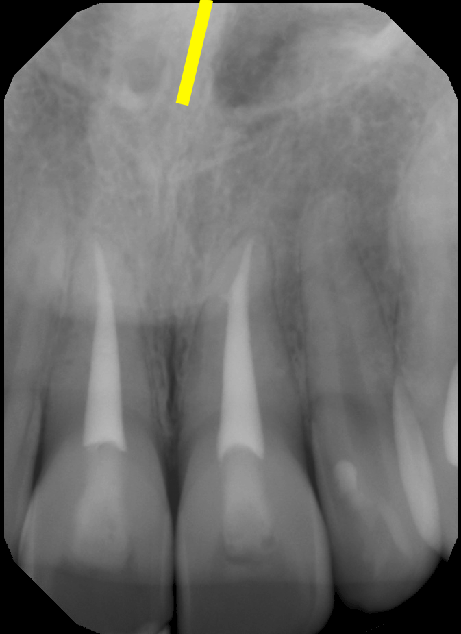
Nasal Septum
The nasal septum appears as a vertical radiopaque band starting at the floor of the nasal cavity. How much of it is visible is dependent on how much of the nasal cavity is captured.
And there’s the anatomy seen on a single periapical radiograph. I will be making more of these types of anatomy posts along with specific anatomy posts mixing 2D and 3D.
If you have any questions or comments, please leave them below. Thanks and enjoy!