This week is a case of an ameloblastoma over a period of six years and an educational video. The radiographs are a little lower quality due to them being duplicates of old radiographs but you can still see the progression over time. This is also a less common location to find an ameloblastoma.
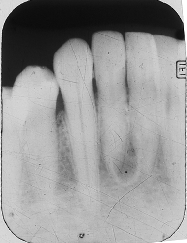
Here is the initial periapical radiograph.
 Note the well-defined corticated radiolucent entity between the apices of the central and lateral incisors.
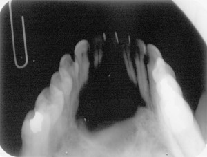
And six years later here is an occlusal radiograph made in the same area.
 Note the (much larger) radiolucent area in the anterior mandible.
Now, onto an educational video made by UNMC College of Dentistry Class of 2016 students about ameloblastoma.


Nice case, Shawneen, which again underlines the importance of an early investigation and diagnosis. Congrats to your students also. Cheers, Marc