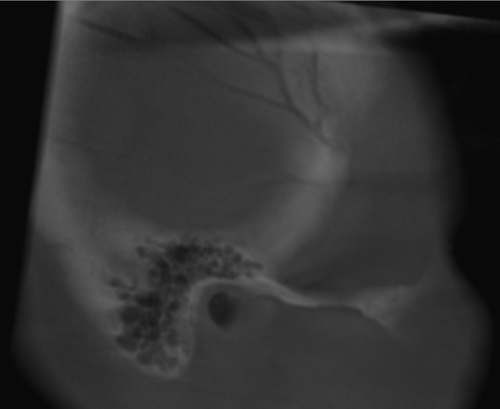
I realize it’s been a while since I’ve posted anatomy so this week I wanted to show a fun case of prominent vascular markings noted in the cranium on a cone beam CT scan. Check it out below.
Note the ‘tree branch’ appearance at the superior aspect of this sagittal view. (This is a 5 mm slice)
If you have any questions or comments, please leave them below. Thanks and enjoy!

As always a good post.
I have a question – unrelated to this post but can you please clarify for me?
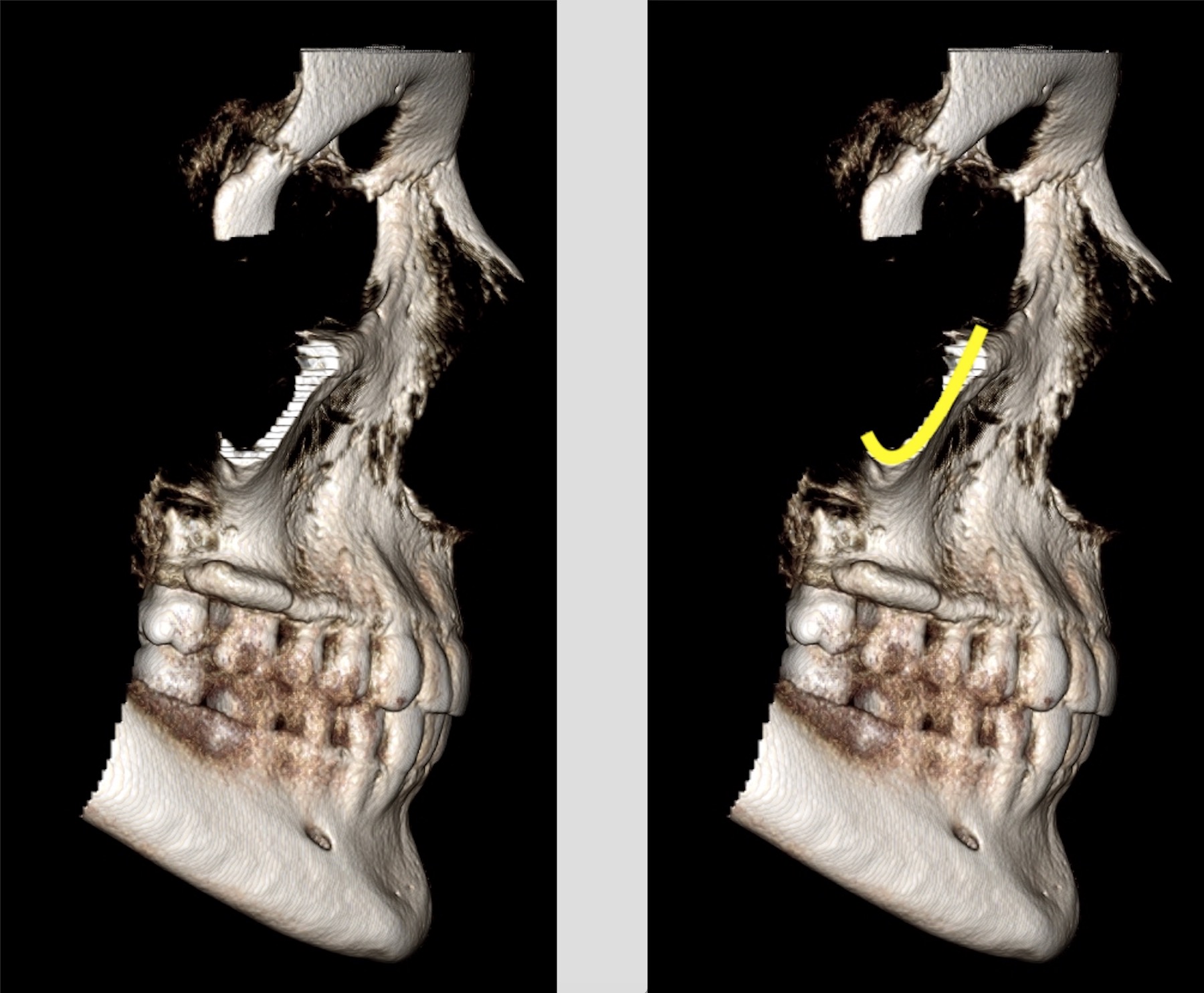
what exactly is the white line in a panoral radiograph. I have always thought it to be the palate ( palata lprocess of the maxilla) producing a white line due to its curvature as the beam rotates around the patient.
I have looked at your OPG anatomy series and I am not sure about this feature?
Thank you for your time.
It sounds as if you are describing the hard palate / floor of the nasal cavity which produces a radiopaque line superior to the maxillary teeth on a pantomograph. You appear to have it correct. 🙂
A question that may not be related to this post
when you see prominent vascular markings in the anterior mandible inter foraminal zone – or the so called incisive canal – how significant is it when u place an implant?
will there be excessive bleeding?
will there be any numbness of the lip area or the lower alveolus?
Based on a few conversations I’ve had with those who have placed implants into this location, I have not heard of any excessive bleeding issues but have heard of a few cases of numbness. I haven’t had a chance to thoroughly review this topic in the research so please take this as my little bit of information I’ve come across regarding this topic. Thanks. 🙂
thank you madam
appreciate your input
is there any report on the incidence of an anterior extension of the IAN
I am not aware of any reports on the incidence but I have not had a chance to do a thorough evaluation of the research out there.
Great post..nice to know about Vascular Markings.