1 min read 0

Anatomy Monday: Pterygomaxillary Fissure

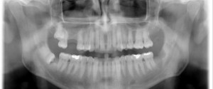
This week I have a few pantomographs showing the pterygomaxillary fissure.  The pterygomaxillary fissure is the lateral opening to the pterygopalatine fossa. The pterygomaxillary fissure presents as a radiolucent inverted teardrop shape just lateral to the maxilla. It has a a radiopaque edge.   If you have any questions about…
1 min read 0

Case of the Week: Mucous Retention Pseudocyst

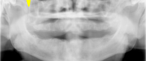
This week I have a case of a mucous retention pseudocyst on a pantomograph. Mucous retention pseudocysts are incidental findings that do not require treatment. They are most commonly found in the maxillary sinuses followed by the sphenoid sinuses. Note the rounded radiopaque dome in the right maxillary sinus. If…
1 min read 0

Anatomy Monday: Incisive Foramen

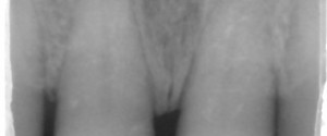
Last week I showed the superior foramina of the nasopalatine canal and this week is the inferior foramen; the incisive foramen.  The incisive foramen presents as a round to ovoid radiolucent entity between the maxillary central incisors.  When the width of the incisive foramen is 10 mm (1 cm) or…
1 min read 6

Locate the Object: September 2013

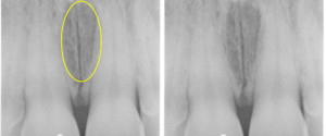
Onto another fun case to use your image shift and SLOB (Same-Lingual, Opposite-Buccal) information. The object in question is the round radiolucent entity seen over the crown of the left central incisor. What surface (buccal or lingual) is it on? The answer will be coming in two weeks (September 24th). …
1 min read 1

Anatomy Monday: Superior Foramina of the Nasopalatine Canal

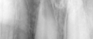
Now that all the foramina of the mandible have been covered (lingual, mental and mandibular) it’s onto the maxilla.  The first one I am going to show is the superior foramina of the nasopalatine canal.  The nasopalatine canal splits superiorly with an opening in the right and left nasal cavities.The…