Skip to content
Dr. G's Toothpix

Dr. G's Toothpix

An oral radiology source for dental professionals.

  • START HERE
  • Radiology Reporting Service
    • Services / Pricing
    • How To Upload A Case
    • Sample Report
    • Contact
  • Describing Radiographic Lesions
  • Radiographic Interpretation
    • Benign Neoplasms
      • Ameloblastoma
      • Cementoblastoma
      • Compound Odontoma
      • Enostosis
      • Focal Idiopathic Osteosclerosis
      • Odontogenic Keratocyst
    • Cysts of the Jaws
      • Buccal Bifurcation Cyst
      • Dentigerous Cyst
      • Lateral Periodontal Cyst
      • Nasopalatine Canal Cyst
      • Periapical / Radicular cyst
      • Simple Bone Cyst
    • Maxillary Sinuses
      • Antral Exostoses / Antral Projection
      • Antrolith
      • Mucositis
      • Mucous Retention Pseudocyst
      • Sinusitis
    • Periapical Inflammatory Diseases
      • Rarefying Osteitis
      • Sclerosing Osteitis
      • Widened Periodontal Ligament Space
    • Skeletal Developmental Abnormalities
      • Cleft Palate
      • Stafne Defect
    • Soft Tissue Calcifications / Ossifications
      • Antrolith
      • Arterial calcifications
      • Ossified Stylohyoid Ligament
      • Osteoma Cutis
      • Rhinolith
      • Sialolith
      • Tonsiliths
    • Tooth anomalies
      • Abrasion
      • Amelogenesis Imperfecta
      • Ankylosis
      • Attrition
      • Dens Evaginatus
      • Dens Invaginatus
      • Dentinogenesis Imperfecta
      • Ectopic Eruption
      • Enamel Pearl
      • External Resorption
      • Gemination
      • Hypercementosis
      • Internal Resorption
      • Macrodont
      • Microdont
      • Oligodontia
      • Pulp Stone
      • Pulpal Sclerosis
      • Supernumerary teeth
      • Talon cusp
      • Taurodontism
      • Transposition
    • Trauma to Teeth and Jaws
      • Coronal fractures
      • Fibrous Scar
      • Mandibular Fractures
      • Root fractures – Horizontal
      • Root fractures – Vertical
  • About
    • Disclaimer
    • Publications

Educational Video: Multiple Myeloma

Posted on July 17, 2013July 10, 2013 by Dr. Shawneen Gonzalez

Another well done educational video by Dental Class of 2015 student David Pelster.

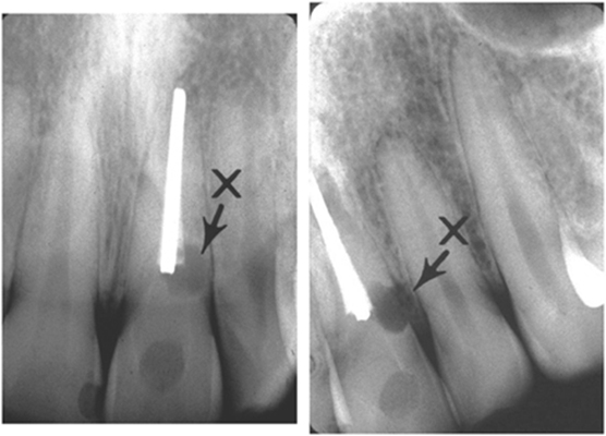
Again, still in search of more cases of this so please let me know if you have a good case you’d like to share. Thanks and enjoy!

Posted in Radiographic InterpretationTagged multiple myeloma, videos

Post navigation

Previous: Locate the Object: July 2013
Next: Case of the Week: Enamel Pearls

Looking for something?

Post topics

anatomy anterior nasal spine artifacts caries caries interpretation CBCT CBCT anatomy CBCT interpretation common radiographic errors cysts of the jaws digital radiography external resorption floor of the nasal cavity image shift impacted tooth intraoral radiograph anatomy intraoral radiographic technique lateral cephalometric skull anatomy LESION lingual foramen locate the object maxillary sinuses mental foramen microdont oral radiology websites pantomograph anatomy pantomograph positioning phosphor plates pulpal/apical findings radiation physics radicular / periapical cyst radiographic interpretation radiographic quality evaluation radiographic technique rarefying osteitis resorption sensors SLOB rule soft tissue calcifications / ossifications supernumerary teeth supernumerary tooth tooth anomalies trauma to teeth and jaws videos x ray

Related Posts

  • Radiographic Interpretation

How was that missed? (Impacted teeth)

  • Dr. Shawneen Gonzalez
  • August 8, 2012
  • 2 min read
  • 6
  • Radiographic Interpretation

Educational Video: Sickle Cell Anemia

  • Dr. Shawneen Gonzalez
  • July 9, 2014
  • 1 min read
  • 0
  • Radiographic Interpretation

Locate the Object: May 2014

  • Dr. Shawneen Gonzalez
  • May 13, 2014
  • 1 min read
  • 3
  • Radiographic Interpretation

Lateral Cephalometric Skull Anatomy – Part V

  • Dr. Shawneen Gonzalez
  • April 17, 2013
  • 1 min read
  • 0
Copyright © 2025 Dr. G's Toothpix Theme: Flawless Blog By Adore Themes.