Sorry to be gone for such a long hiatus. I’ve been working on a side project that has taken much more time than expected. If all goes well I’ll be posting in a few months the outcome of that project. Now onto some anatomy. 🙂
The mental foramen is most commonly seen near the apex of the mandibular second premolar. It may be visible anteriorly to the first premolar and posteriorly to the first molar. It presents as a radiolucent round to ovoid entity. When it is superimposed over the apex of a tooth it is important to look at the periodontal ligament space and lamina dura of the tooth to determine a pathosis of tooth origin versus the mental foramen.
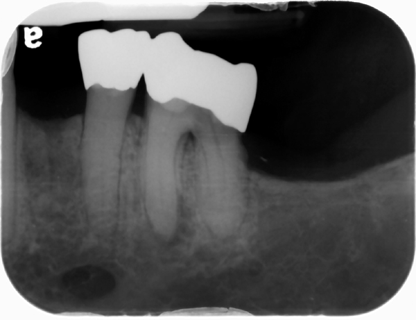 Periapical radiograph showing mental foramen as well-defined, ovoid radiolucent area apical to second premolar.
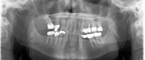
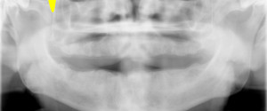
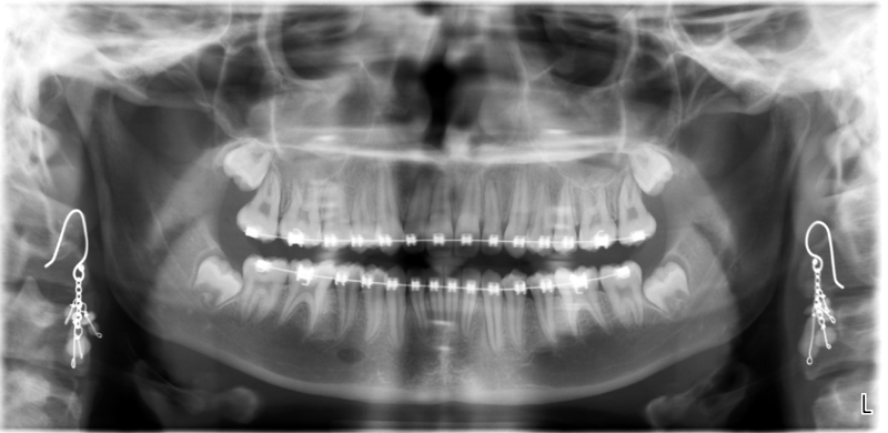 Pantomograph showing right mental foramen as well-defined, ovoid radiolucent area apical to mandibular premolars.
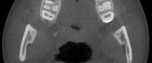
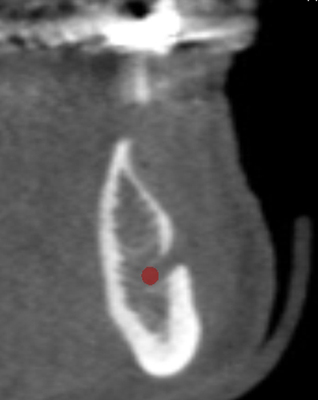 Cone Beam CT cross-sectional slice showing opening of mental foramen as a discontinuity of the facial cortical plate. Mandibular canal noted by red circle.
If you have any questions or would like to see other examples, please let me know and I’ll see what I can. Thanks and enjoy!

sir please let me know the syndromes associated with anomalies with mental foramen
I am not sure what kind of anomalies you are wanting to know about with the mental foramen – absence of it, double foramina, etc. Please let me know. Thanks.