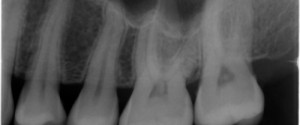
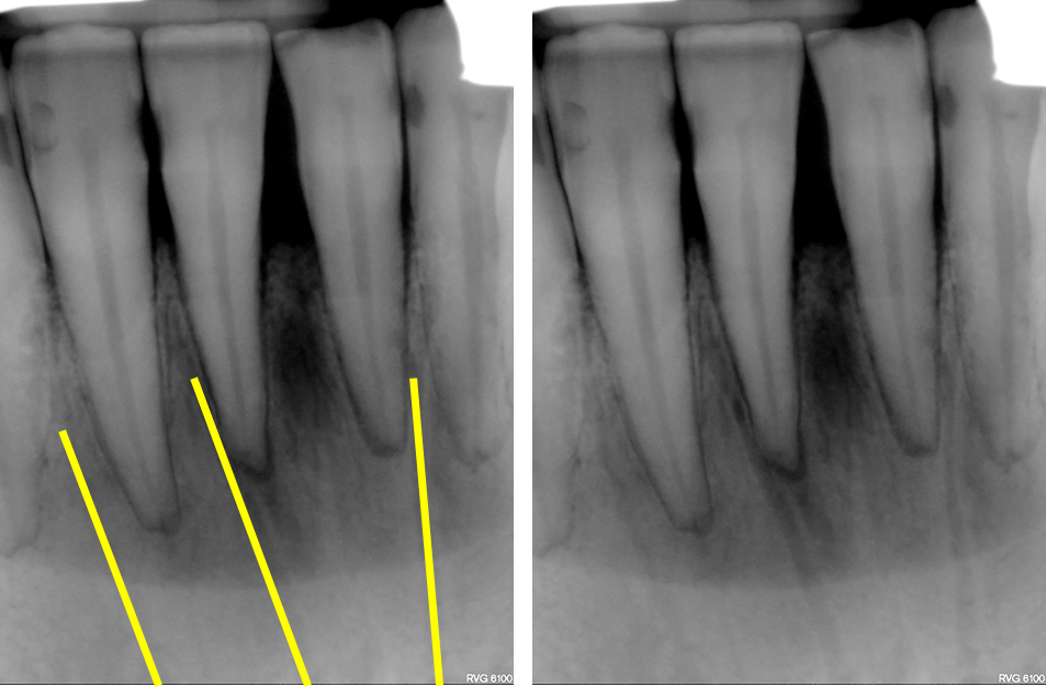
Today I am starting a new series for Mondays on anatomy. I will be showing different anatomical landmarks for both intraoral and extraoral radiographs. This first entity I am showing is a radiographic anatomical landmark; the Y line of Ennis. This is sometimes referred to as an Inverted Y. It is created by the superimposition of the floor of the nasal cavity (straight radiopaque line) and the border of the maxillary sinus (curved radiopaque line).
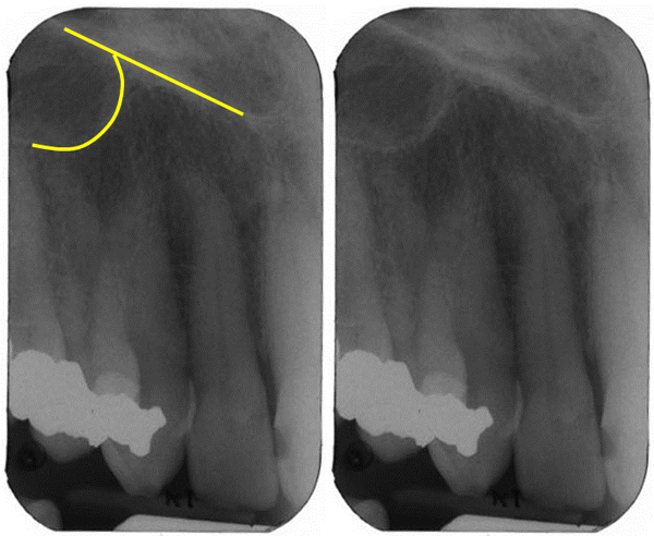 Y line of Ennis (yellow lines)
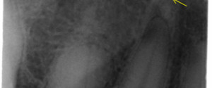
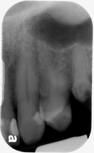 Y line of Ennis and palatal tori (radiopaque mass distal)
If you have any questions or comments or would like to see a specific anatomical entity, please leave them below. Thanks and enjoy!
SPONSOR

Nice!
Any specific tooth region related to this line??
The maxillary canine and first premolar are typically seen in the Y line of Ennis region.
In which view we can see this Y line sir?
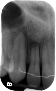
Both images show the Y line of Ennis or are you asking typically which specific radiographs show this landmark? If the latter, its typically seen on maxillary canine periapical radiographs and sometimes maxillary premolar periapical radiographs.
Thank you!