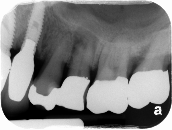
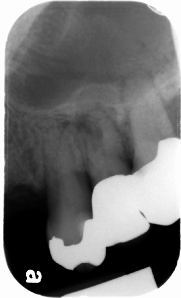
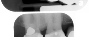
This months how was that missed is a case of chronic apical periodontitis or rarefying osteitis. The first radiograph is 2 years prior to the second one.
Initial interpretation: No initial interpretation as this radiograph was made for an implant in the maxillary canine region and nothing else was evaluated. 🙁
Final Interpretation: Rarefying osteitis at the apex of the maxillary left second premolar with external resorption (most likely due to the presence of a periapical granuloma at some time). Upon reviewing the older radiographs, the rarefying osteitis was evident on radiographs from 2 years prior.
Lesson learned: It is important to remember to evaluate the entire radiograph and not focus on only the area or tooth where treatment is planned. This case is a good reminder that every aspect of every radiograph must be thoroughly evaluated.
If you have any questions about this case, please let me know. Thanks and enjoy!
Next week: Pantomograph Positioning Errors Part 2
SPONSOR

is tht cause roluscent at max left second premolar from heavy contact?is it possible of tht?bcse if it was from previous granuloma it means tht tooth nonvtal n shuold be treated wth endodontic,but we ddnt see any endo treatmen…thks for discussion