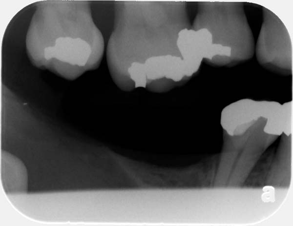
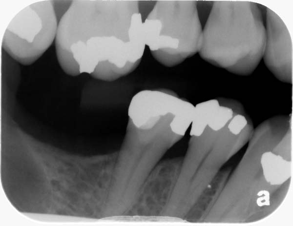
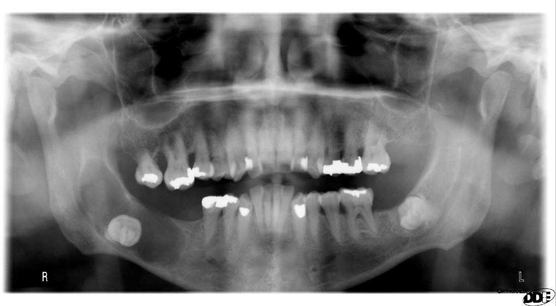
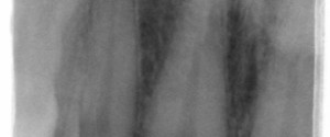
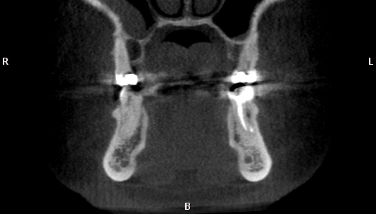
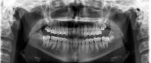
This week is a case of a quite large dentigerous cyst. A dentigerous cyst will appear radiolucent surrounding the crown of an impacted tooth. The teeth most commonly effected are the maxillary canines and third molars (both maxilla and mandible). A pathognomonic sign of a dentigerous cyst is that the border of the lesion is continuous with the cemento-enamel junction of the impacted tooth. This case was first identified on bitewing radiographs noting a loss of bone with a radiopaque mass (impacted third molar) in this area. A pantomograph was ordered to further evaluate the area. The pantomograph shows a well-defined radiolucent entity surrounding the crown of the impacted right mandibular third molar. The patient was referred to an oral surgeon for excision of the lesion.
Bitewing radiographs showing radiolucent area with radiopaque mass (third molar) in the mandible
Pantomograph
For more information and other radiographs of dentigerous cysts check out the page on dentigerous cysts.
Enjoy!
