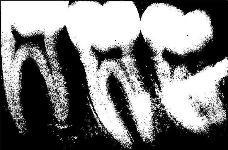
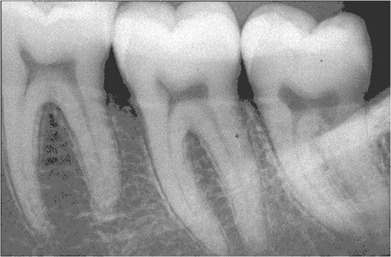
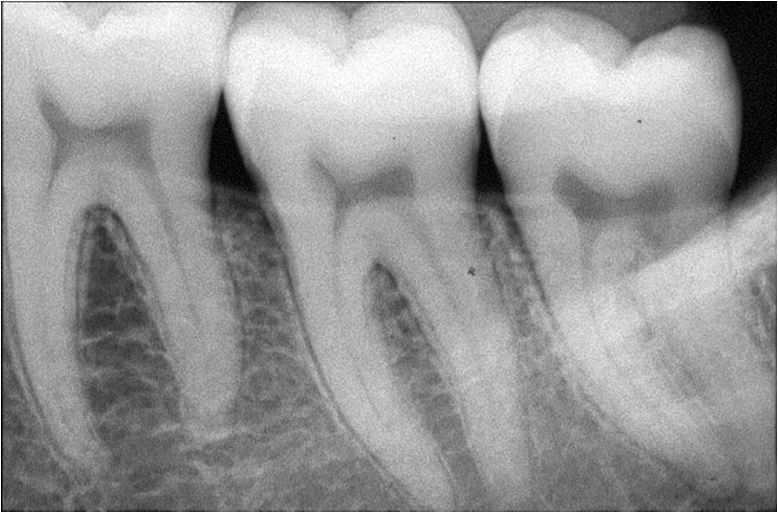
This weeks topic on digital radiography is contrast resolution. Contrast resolution is the ability to distinguish gray levels on an image. The term used to identify how many gray levels that can be identified is bits. When any number is in front of bits, such as 8 bits or 14 bits, that number refers to a power of the number 2. So if an image has 8 bits, there is 2 to the power of 8 ( which is 2 x 2 x 2 x 2 x 2 x 2 x 2 x 2 or 256 gray levels possible). As the number increases, so does the number of gray levels possible in an image. Below are sample images with different bit depths. (Images courtesy Dr. Noujeim – UTHSCSA)
While increasing bit depth increases the overall number of grays in an image, this does not necessarily improve interpretation of radiographs. The biggest roadblock is conventional monitors. A conventional monitor (any monitor that comes standard with a computer, laptop, etc.) is only 8 bits. So if you have 14 bit sensor or phosphor plate and the information is captured on the computer, you can only see 256 grays. It is possible to buy a higher resolution monitor. These are typically radiology monitors purchased from medical companies. These monitors are referred to in relation to megapixels, as in 3 megapixels, 5 megapixels and so on. These monitors are 12 bit monitors and can show up 4096 gray levels. These monitors are pretty expensive with lower tier models (3 megapixels) starting around ~$8,000 (US dollars).
Other issues to consider is lighting. When viewing radiographs in a dimmed room with excess light blocked, the human eye can identify up to 60 different gray levels. If the room is not dimmed (typical of a dental operatory) the number of gray levels visible falls to 30.
I hope this little information about contrast resolution and digital radiography is helpful. If you have any questions or comments, please let me know.
Thanks and enjoy!


Dear Dr. Shawneen Gonzalez
How I can get the permission to published as an attached to my PH.D. thesis the following figure from Digital Radiography: Contrast resolution 1to 8 bit intraoral images (Images courtesy Dr. Noujeim – UTHSCSA) ?
My thesis ( Effect of display type and room illuminance in digital dental radiographs. -Display performance in panoramic and intraoral radiography, 2015) will be published in the series Acta Universitas Ouluensis of the University of Oulu.
Yours sincerely
Soili Kallio-Pulkkinen
DDS
Department of Diagnostic Radiology
Oulu University Hospital
P.O.Box 50
Fin-90029 Oulu University Hospital
Finland
E-mail: soili.kallio-pulkkinen@oulu.fi
soili.kallio-pulkkinen@ppshp.fi
Tel. +3583153847
Soili,
Being that those images are not mine you would need to reach out directly to Dr. Noujeim at UTHSCSA for permission. If you look at the dental school in San Antonio you will be able to find his contact information.
Thanks,
Shawneen