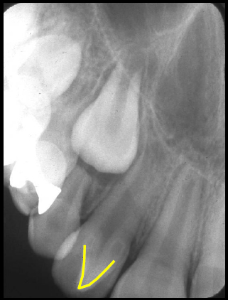
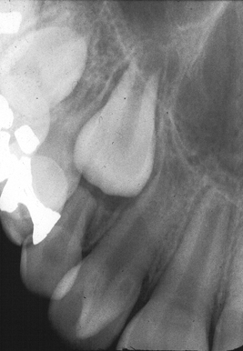
Definition: Hyperplasia of the cingulum of an anterior tooth.
Radiographic Features:
Location: Any anterior tooth (primary or permanent dentition).
Edge: Well-defined.
Shape: Triangular, superimposed over crown of associated tooth.
Internal: Radiopaque, same radiopacity as enamel.
Other: None.
Number: Can be either single or multiple.
(click image to enlarge)
Talon cusp
(left – note yellow line over cusp) (right – note triangular radiopacity)


I have a talon cusp on my right maxillary lateral incisor..do u think research could be done for educational purposes..I’d love to further dental knowldge.
It would depend on the specific talon cusp and if it’s outside the range of normal. Talon cusps have been heavily researched in the past so only abnormal cases would likely be added to the literature.