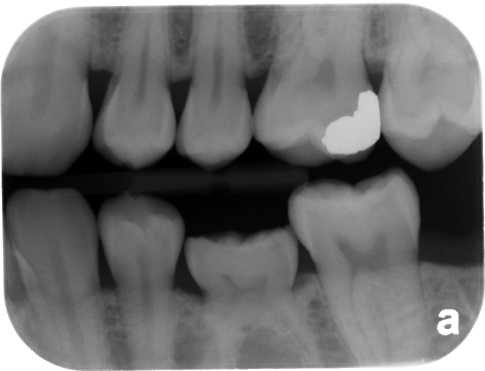
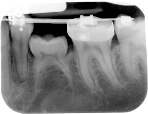
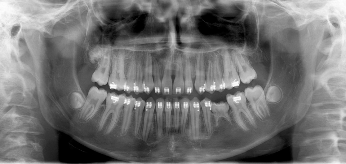
Definition: The fusion of a tooth root with the surrounding bone due to an absence of the periodontal ligament space.
Radiographic Features:
Location: Any tooth in the oral cavity, frequently associated with primary second molars.
Edge: Well-defined, normal outline of a tooth.
Shape: Toothlike.
Internal: Radiopaque, same radiopacity as tooth structure.
Other: There will be a step in the occlusal plane where the ankylosed tooth is present in some cases. Beware of the ‘absent periodontal ligament space’ meaning ankylosis. This is more likely due to radiographic technique.
Number: Usually single, but may be multiple.
Ankylosis
(note the step in the occlusal plane with the left mandibular primary second molar)
Ankylosis
(note the step again in the occlusal plane)
Ankylosis
(left mandibular primary second molar with step in the occlusal plane)


One thought on “Ankylosis”