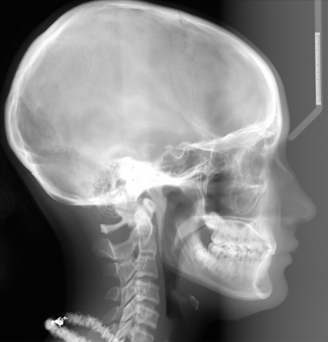
I had a request a couple months ago wanting more anatomy on lateral cephalometric skull radiographs specifically those landmarks used in orthodontics. As I am not an orthodontist and do not make tracings of lateral cephalometric skull I will not be going over how to trace but where the anatomical landmarks are along with radiographic examples.
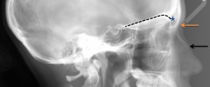
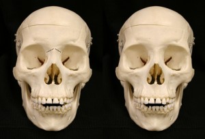
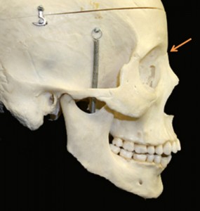
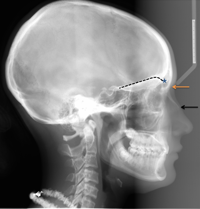
Nasion – the suture between the frontal bone and nasal bones (black dotted line and orange arrow).
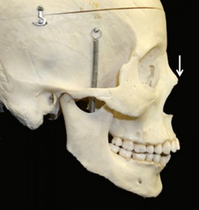
Rhinion – tip of the nasal bone (white arrow).
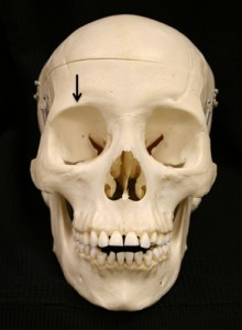
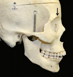
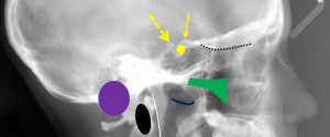
Suprarobital – junction of the orbit at the superior aspect where it becomes the roof of the orbital cavity (black arrows).
Roof of the orbital cavity – just like the name implies; the roof of the orbital cavity.
Rhinion (black arrow)
Supraorbitale (blue star)
Roof of the orbital cavity (black dotted line)
I’m not sure how many parts this will take but it’ll be a few so more coming. If you have any questions or comments, please leave them below. Thanks and enjoy!
One thought on “Lateral Cephalometric Skull Anatomy – Part I”
Comments are closed.