1 min read 0

Educational Video: Sinusitis

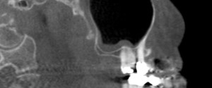
This week I have a case of sinusitis on cone beam CT along with another educational video. 🙂 On the cone beam CT images note the radiopaque band that follows the floor and the posterior border on the sagittal view and the floor and medial border of the left maxillary…
1 min read 0

Case of the Week: Palatal Tori

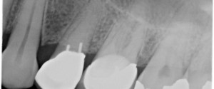
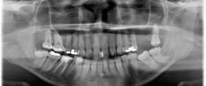
This week I have a case of palatal tori evident not only on a periapical radiograph but a pantomograph as well.  On the periapical radiograph it appears as a radiopaque mass superior to the maxillary teeth. The pantomograph shows a well-defined horizontal radiopaque band directly inferior to the floor of…
1 min read 4

Educational Video: Attrition

This week I have a case of attrition along with an educational video.  First the case; this is an example of attrition on the lingual surfaces of the maxillary incisors.  It presents with the crown having an increased radiolucent appearance and a defined horizontal line near the cemento-enamel junction where…
1 min read 0

Case of the Week: Talon Cusp

This week I have a case of a talon cusp so large it’s evident on a pantomograph.  While there is more than one talon cusp evident on this radiograph, it’s the one on the maxillary left lateral incisor that is nearly as big as the crown that’s amazing :).  Note…