1 min read 0

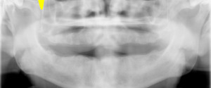
Anatomy Monday: Pterygomaxillary Fissure

This week I have a few pantomographs showing the pterygomaxillary fissure.  The pterygomaxillary fissure is the lateral opening to the pterygopalatine fossa. The pterygomaxillary fissure presents as a radiolucent inverted teardrop shape just lateral to the maxilla. It has a a radiopaque edge.   If you have any questions about…
1 min read 8

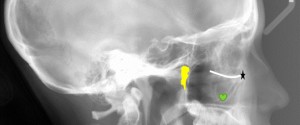
Lateral Cephalometric Skull Anatomy – Part II

Onto part 2 of lateral cephalometric skull anatomy frequently used in orthodontics. Again identified on a dry skull first followed by the radiograph. Orbitale (black star) – The lowest point of the orbital margin (also the anterior point for the Frankfort horizontal plane). Key ridge (green heart) – The inferior…
2 min read 10

Anatomy on Radiographs: Pantomographs Part 2

Now onto other structures visible on a pantomograph. Midface The orbital rim (green dots) appears as a thick semi –circular radiopaque band superior to the maxillary sinuses. The pterygomaxillary fissure (blue area) appears as an inverted teardrop shaped radiolucent area with a thin radiopaque border. It is directly lateral to…