1 min read
0
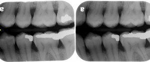
Radiographic Quality Evaluation: Posterior Bitewing Radiographs
Now onto evaluation of bitewing radiographs and what to look for. 1. The teeth to be captured are recorded on the radiograph. This means that no portion the crown is cut off on the radiograph. 2. A minimum of 2 mm of alveolar crest in both the maxilla and mandible…