1 min read 0

Anatomy Monday: Mandibular Foramen

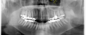
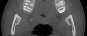
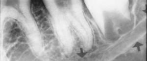
Since I just finished a series of anatomy post of canals I find it fitting to now move onto foramina. 🙂  I’ve already covered a few in the past; the lingual foramen and the mental foramen.  So I will finish off the mandible with today’s post of the mandibular foramen.…
1 min read 0

Anatomy Monday: Infraorbital Canal

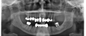
The infraorbital canal is seen on extraoral radiographs specifically pantomographs.  It appears as a diagonal radiolucent band with two thin radiopaque borders superimposed over the inferior border of the orbital rim. If you have any questions or comments about the infraorbital canal and its appearance on radiographs, please leave them…
1 min read 0

Anatomy Monday: Nasopalatine Canal

It’s time for the next canal: the nasopalatine canal.  This canal may also be referred to as the incisive canal.  It is seen on both intraoral radiographs and extraoral radiographs.  The nasopalatine canal presents as a vertical radiolucent band between the roots of the maxillary central incisors superiorly to the…
1 min read 0

Anatomy Monday: Mandibular Canal

This week and month is going to be canals and ducts visible on intraoral and extraoral radiographs. I’m starting with a canal frequently sought out specifically for implants and extractions; the mandibular canal.  The mandibular canal is visible on both intraoral radiographs and extraoral radiographs. It presents as a radiolucent…
1 min read 3

Anatomy Monday: Nasolabial Fold (soft tissue)

This week is another soft tissue entity that can be seen on both intraoral and extraoral radiographs. The nasolabial fold presents as a diagonal transition line. A transition line is seen as a defined line where part of the radiograph appears more radiopaque due to superimposition of soft tissue. The…