1 min read 2

Anatomy Monday: Pineal Gland Calcification

This week I’m onto a calcification encountered in the soft tissue of the brain; the pineal gland.  It appears as a well-defined radiopaque entity in the mid-line of the brain superior to the spinal column. Another entity from my book (Interpretation Basics of Cone Beam Computed Tomography). If you have…
1 min read 0

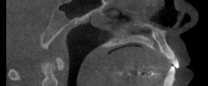
Anatomy Monday: Spheno-Occipital Synchondrosis

Onto another fun entity shown in my new book (Interpretation Basics of Computed Tomography); the spheno-occipital synchondrosis. The spheno-occipital synchondrosis is a normal developmental entity seen as a well-defined discontinuity of the sphenoid bone in children and teens.   If you have any questions or comments about the spheno-occipital synchondrosis,…
1 min read 0

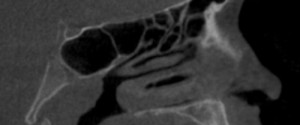
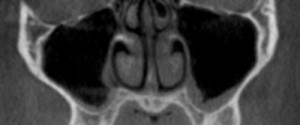
Anatomy Monday: Concha Bullosa

I’ve decided to mix up the Anatomy Monday a little this month celebrating the release of my book – Interpretation Basics of Cone Beam Computed Tomography 😀 by covering entities shown in the book. Today is concha bullosa, an aerated concha most commonly associated with the middle concha. It is…
1 min read 0

Anatomy Monday: Top anatomy of 2013

Here are the most viewed Anatomy Monday posts of 2013. 5. Anterior nasal spine 4. Superior foramina of the nasopalatine canal 3. Nasolabial fold 2. Mental foramen 1. Incisive foramen If there are any other anatomical entities you’d like to see covered in the upcoming posts, please leave a comment…
1 min read 2

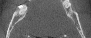
Case of the Week: Tonsiliths

This week is another CBCT case of calcifications, this time in the tonsils.  On CBCT images, these are seen on the axial views lateral to the airway and on coronal views medial to the ramus of the mandible.  They are well-defined irregular shaped radiopaque entities. They may be single or…