1 min read 0

Anatomy Monday: Zygomatic Process of the Maxilla

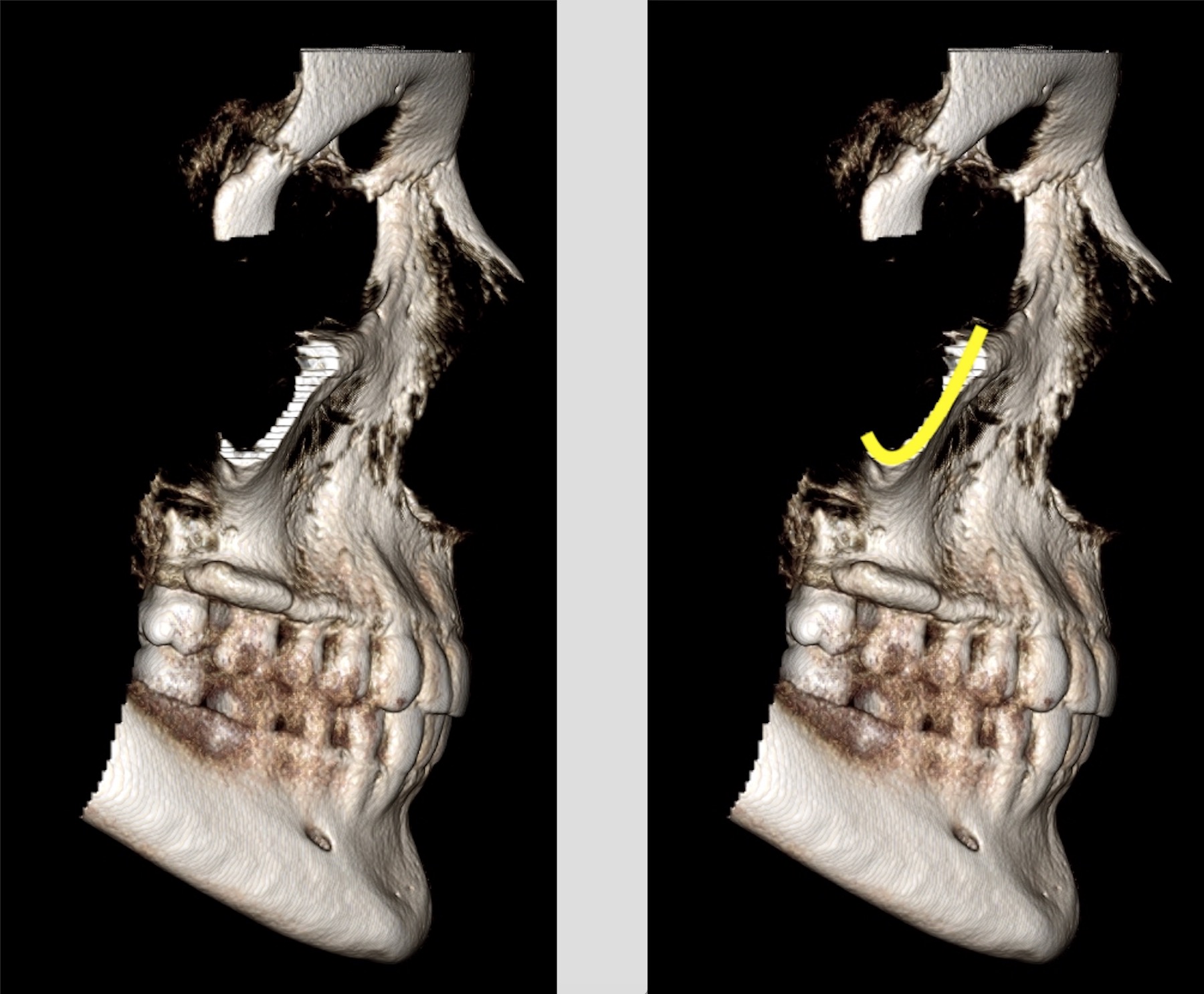
This anatomy can be tricky for some to visualize as its appearance is understood by knowing the direction of x rays and how they course through this process. So to try and show that I’m adding some 3D renderings from a CBCT scan. The zygomatic process of the maxilla appears…
1 min read 0

Lateral Cephalometric Skull Anatomy – Part V

Today is all about the mandible. Condyle (black curved dotted line) Coronoid process (white triangular dotted line) Mandibular first molar (green oval) Infradentale (yellow dotted arrow) – Superior facial bone height near the level of the cemento-enamel junction of the mandibular incisors. B point (white solid arrow) – The posteriormost…
1 min read 1

Anatomy Monday: Floor of the nasal cavity (maxilla)

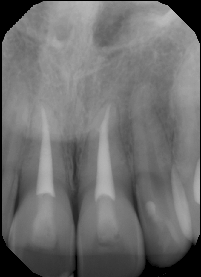
The floor of the nasal cavity that is visible on radiographs is actually the junction of the floor and lateral wall or vomer.  On intraoral radiographs this will appear as a single radiopaque straight line superior to the maxillary teeth.  It is visible on any periapical radiograph (anterior and posterior). …
1 min read 6

Anatomy Monday: Y line of Ennis (maxilla)

Today I am starting a new series for Mondays on anatomy. I will be showing different anatomical landmarks for both intraoral and extraoral radiographs.  This first entity I am showing is a radiographic anatomical landmark; the Y line of Ennis.  This is sometimes referred to as an Inverted Y.  It…
1 min read 4

Case of the week: Double Lingual Foramina

This weeks case is an interesting variant of normal anatomy – two lingual foramina seen on a periapical radiograph.  Some patients can have up to three lingual foramina. Note the two well-defined, circular radiolucent entities inferior to the mandibular central incisors.  The superior one is larger than the inferior one.…