Definition: Ossification of the stylohyoid ligament.
Radiographic Features:
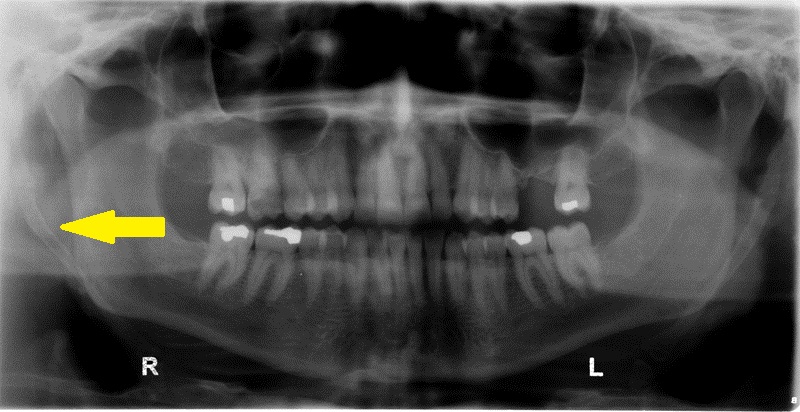
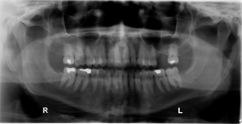
Location: Lateral of the ramus on a pantomograph. Sometimes it may be superimposed over the distal aspect of the ramus.
Edge: Well-defined.
Shape: Linear
Internal: Radiopaque.
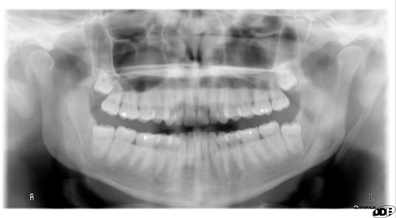
Other: There are multiple ossification centers from which the ligament begins ossification. The areas where opposing ligament ossifications meet will have a ‘joint-like’ appearance of two bones articulating.
Number: Maybe unilateral or bilateral.
TIP: When a patient presents with ossification and PAIN when rotating the head, Eagle’s syndrome must be considered. If a patient has ossified stylohyoid ligament/s but no pain, it is not Eagle’s syndrome.
Ossified stylohyoid ligament
(bilateral with arrow)
Ossified stylohyoid ligament
(bilateral without arrow)
Ossified stylohyoid ligament – Left
(note the ‘joint-like’ appearance on both sides where the two ossification segments meet)


One thought on “Ossified Stylohyoid Ligament”