Definition: A retained root fragment of a deciduous (primary) tooth that has not been resorbed within the mandible or maxilla. The most commonly affected tooth is the deciduous second molar.
Radiographic Features:
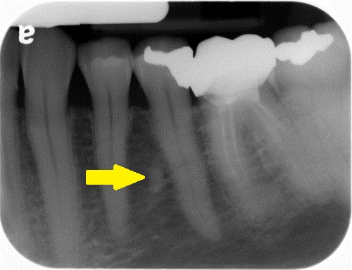
Location: Most commonly seen on the mesial and/or distal of the second premolar. The mandible is more common than the maxilla. There can be retained deciduous root fragments of other deciduous teeth seen throughout the maxilla and mandible.
Edge: Well-defined. There may still be evidence of a periodontal ligament space around the root fragment.
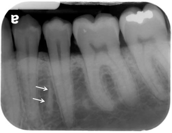
Shape: Frequently to be linear or curved.
Internal: Radiopaque, radiopacity of tooth structure specifically dentin.
Other: None.
Number: May be single or multiple.
TIP: If there is no evident periodontal ligament space around the retained root fragment, it can be difficult to determine the difference between an enostosis (dense bone island) or a retained deciduous root fragment. If the radiopaque area has a linear shape, a retained root fragment is most likely. Neither entity needs treatment as they are both incidental findings.
(Click image to enlarge)
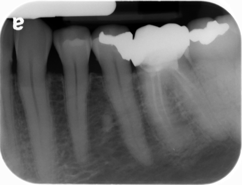
Retained deciduous root fragment
(left – with arrow – note evident periodontal ligament space) (right – without arrow)
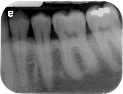
Retained deciduous root fragment
(left – with arrow) (right – without arrow)
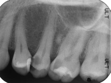
Retained deciduous root fragment
(distal to maxillary left second premolar – # 13)
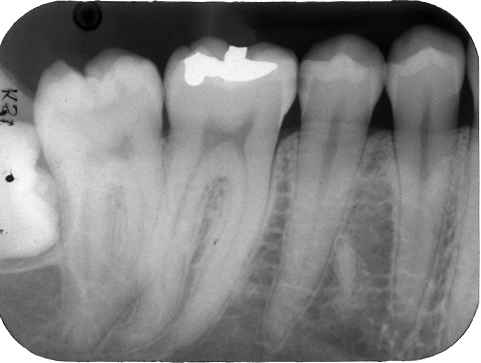
Retained deciduous root fragment
(mesial to mandibular right second premolar – # 29)
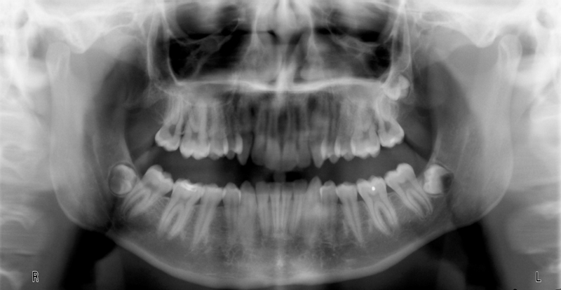
Retained deciduous root fragments
(mesial and distal of bilateral mandibular second premolars – # 20 and # 29)
(note the linear shape)
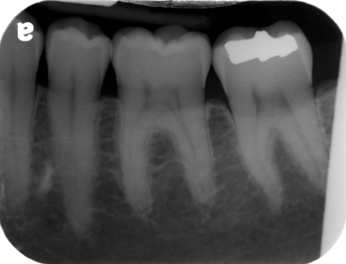
Retained deciduous root fragment
(mesial to mandibular left second premolar – # 20)
I’m excited I found this website. Very helpful. I’m a DDS currently entering a residency program in radiology and hopefully looking to take this specialty to the next level. Keep up the great work!
Thanks! Good luck in your residency and I’m sure our paths will cross in the future. 🙂
could you please explain the differences of apical abcess and apical periodontitis, also whether you can tell the difference if it is acute or chronic in an xray? Thank you in advance.
I will work on showing some examples of the entities you have listed in the upcoming case of the week this month. Thanks.