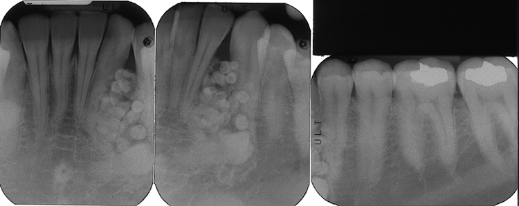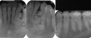
Definition: A tumor consisting of enamel, dentin, cementum and pulp tissue. A compound odontoma will have defined tooth-like shaped entities (denticles) within the lesion.
Radiographic Features:
Location: Maxillary anterior region is most common.
Edge: Well-defined. Corticated.
Shape: Tooth-like shape radiopacities within the lesion.
Internal: Mixed radiolucent/radiopaque with radiopacity of tooth structure (enamel and dentin).
Other: Commonly prevents normal eruption of permanent teeth.
Number: Single. Multiple is rare but possible.
(Click image to enlarge)