Definition: The ‘floor of the nasal cavity’ as labeled on radiographs is actually the junction of the floor of the nasal cavity with the lateral wall and/or vomer.
Radiographic Features:
Location: Superior to the maxillary teeth both anterior and posterior.
Edge: Well-defined, corticated.
Shape: Straight line.
Internal: Radiopaque.
Other: None.
Number: Intraoral radiographs – one. Extraoral radiographs – one, two or three possibly seen.
(click to enlarge)
Floor of the nasal cavity
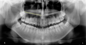
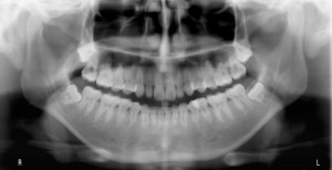 Pantomograph with a single floor of nasal cavity visible on both sides (yellow dotted line)
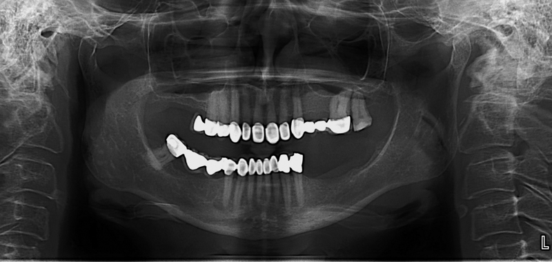 Pantomograph showing two radiopaque lines on patients right.
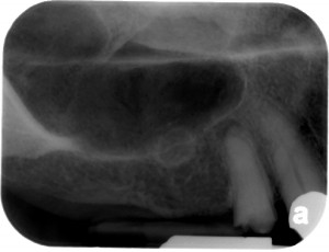
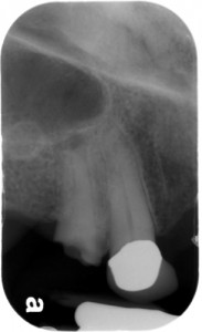
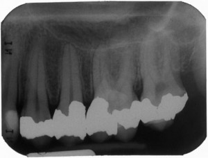
Periapical radiographs
SPONSOR
whats its function and clinical significance? ty
I would have to refer you to an anatomy instructor as I don’t know beyond being the separation of the floor and the oral cavity. 🙂