3 min read 0

Locate the Object: July 2013 Answers

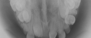
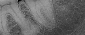
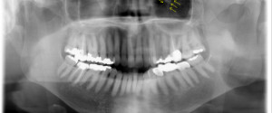
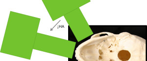
Now onto the answers for the July 2013 Locate the Object.  I will be going over both image shift and SLOB (Same-Lingual, Opposite-Buccal) to determine the location of the pin superimposed over the first premolar. Image shift Before starting to use the image shift principle it is important to know…
1 min read 0

Anatomy Monday: Nasolacrimal Canal

The nasolacrimal duct is most commonly seen on occlusal radiographs specifically the anterior maxillary occlusal radiograph and standard maxillary occlusal radiograph.  It presents as an ovoid radiolucent area with a thin radiopaque border at the lateral edge of the nasal cavity in the region of the molars. If you have…
1 min read 0

Case of the Week: Hypercementosis

This week is a fun case of hypercementosis at the apex of a molar root.  Note on the distal root of the first molar a radiopaque mass with a thin radiolucent line around it. This shows that the mass is attached to the root surface with the periodontal ligament space…
1 min read 0

Anatomy Monday: Infraorbital Canal

The infraorbital canal is seen on extraoral radiographs specifically pantomographs.  It appears as a diagonal radiolucent band with two thin radiopaque borders superimposed over the inferior border of the orbital rim. If you have any questions or comments about the infraorbital canal and its appearance on radiographs, please leave them…