1 min read 0

Lateral Cephalometric Skull Anatomy – Part V

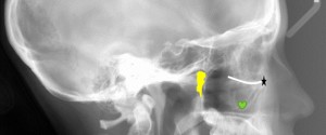
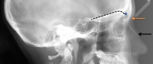
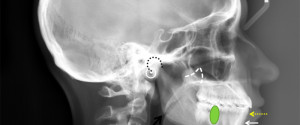
Today is all about the mandible. Condyle (black curved dotted line) Coronoid process (white triangular dotted line) Mandibular first molar (green oval) Infradentale (yellow dotted arrow) – Superior facial bone height near the level of the cemento-enamel junction of the mandibular incisors. B point (white solid arrow) – The posteriormost…
1 min read 0

Lateral Cephalometric Skull Anatomy – Part IV

This post will be all about the maxilla. Anterior nasal spine (yellow arrow) – anterior-most point where right and left maxilla meet on the midline forming a protruding bone spicule. A point (white arrow) – the deepest point between the anterior nasal spine and prosthion. Prosthion (yellow dotted arrow) –…
1 min read 0

Lateral Cephalometric Skull Anatomy – Part III

Now onto the temporal region. Zygomatic Arch (green area) – Made up by the zygomatic bone and zygomatic process of the temporal bone. Mandibular Notch (blue line) – Notch between the condyle and coronoid process of the mandible. Posterior margin of the ramus (white line) – Just as the name…
1 min read 8

Lateral Cephalometric Skull Anatomy – Part II

Onto part 2 of lateral cephalometric skull anatomy frequently used in orthodontics. Again identified on a dry skull first followed by the radiograph. Orbitale (black star) – The lowest point of the orbital margin (also the anterior point for the Frankfort horizontal plane). Key ridge (green heart) – The inferior…
1 min read 1

Lateral Cephalometric Skull Anatomy – Part I

I had a request a couple months ago wanting more anatomy on lateral cephalometric skull radiographs specifically those landmarks used in orthodontics. As I am not an orthodontist and do not make tracings of lateral cephalometric skull I will not be going over how to trace but where the anatomical…