1 min read 0

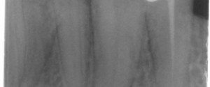
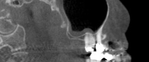
Educational Video: Sinusitis

This week I have a case of sinusitis on cone beam CT along with another educational video. 🙂 On the cone beam CT images note the radiopaque band that follows the floor and the posterior border on the sagittal view and the floor and medial border of the left maxillary…
1 min read 3

Locate the Object: August 2013

Onto another fun case to use your image shift and SLOB (Same-Lingual, Opposite-Buccal) information. The object in question is the left lateral incisor. Where is it (buccal or lingual) in relation to the left central incisor? The answer will be coming in two weeks (August 27th).  You can answer either…
1 min read 4

Educational Video: Attrition

This week I have a case of attrition along with an educational video.  First the case; this is an example of attrition on the lingual surfaces of the maxillary incisors.  It presents with the crown having an increased radiolucent appearance and a defined horizontal line near the cemento-enamel junction where…
3 min read 0

Locate the Object: July 2013 Answers

Now onto the answers for the July 2013 Locate the Object.  I will be going over both image shift and SLOB (Same-Lingual, Opposite-Buccal) to determine the location of the pin superimposed over the first premolar. Image shift Before starting to use the image shift principle it is important to know…