1 min read 0

Anatomy Monday: Paradoxic Curvature

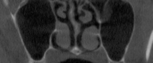
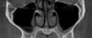
This week is the last anatomy I’ll be showing from my book (Interpretation Basics of Cone Beam Computed Tomography).  It’s another aspect of the nasal cavity involving the middle concha. Paradoxic curvature is when the curve of the concha is towards the nasal septum instead of the lateral border of…
1 min read 2

Anatomy Monday: Pineal Gland Calcification

This week I’m onto a calcification encountered in the soft tissue of the brain; the pineal gland.  It appears as a well-defined radiopaque entity in the mid-line of the brain superior to the spinal column. Another entity from my book (Interpretation Basics of Cone Beam Computed Tomography). If you have…
1 min read 0

Anatomy Monday: Spheno-Occipital Synchondrosis

Onto another fun entity shown in my new book (Interpretation Basics of Computed Tomography); the spheno-occipital synchondrosis. The spheno-occipital synchondrosis is a normal developmental entity seen as a well-defined discontinuity of the sphenoid bone in children and teens.   If you have any questions or comments about the spheno-occipital synchondrosis,…
1 min read 0

Anatomy Monday: Concha Bullosa

I’ve decided to mix up the Anatomy Monday a little this month celebrating the release of my book – Interpretation Basics of Cone Beam Computed Tomography 😀 by covering entities shown in the book. Today is concha bullosa, an aerated concha most commonly associated with the middle concha. It is…
3 min read 3

Article Review: CBCT (JADA August 2012)

There was a recent article in the Journal of American Dental Association (JADA) August 2012 regarding the use of cone beam CT in dentistry.  I thought I would summarize it here as well as give a link to the article itself here.  The article was written by the Council on…