1 min read 2

Case of the Week: Synovial Chondromatosis

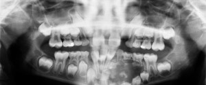
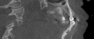
This week is another case and educational video (made by UNMC College of Dentistry Class of 2016 Dental Students). Synovial chondromatosis is uncommon but here is a bilateral case seen on a pantomograph. Note the multiple radiopaque entities anterior to the condyles. Now for more goodies about this entity with…
1 min read 0

Case of the Week: Osteopetrosis

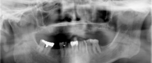
Case and educational video time. 🙂 On this radiograph, note the increased radiopacity of the mandible making it appear completely radiopaque with no bone trabeculation evident. Also note the increased size of the mandible. Now onto the another fun educational video made by UNMC College of Dentistry Class of 2016…
1 min read 2

Case of the Week: Ameloblastic fibro-odontoma

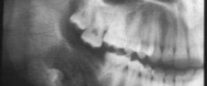
This week is a case and a video. The case is a little older hence it was given to me chopped off (partial radiograph only). I’m pretty sure you can find the area of the ameloblastic fibro-odontoma. Now onto the educational video to learn more about ameloblastic fibro-odontoma made by…
1 min read 0

Case of the Week: Adenomatoid Odontogenic Tumor (AOT)

This week I have a case of adenomatoid odontogenic tumor (AOT) and another educational video.  This case shows the initial tumor and a post op radiograph of several years later with healing. Adenomatoid odontogenic tumors frequently appear radiographically similar to dentigerous cysts encompassing the crown of an unerupted tooth. Now,…
1 min read 0

Case of the Week: Odontogenic Myxoma

This week is another case and video.  This case shows a well-localized radiolucent area between the mandibular left first premolar and canine on the reconstructed pantomograph. It is causing displacement of the roots of these teeth. The axial and sagittal views show thinning with displacement of the facial cortical plate…