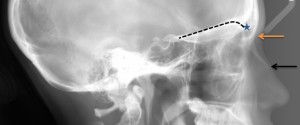
1 min read 0

Top posts and pages of 2012

Here are the top 5 posts and pages of 2012. Posts 5. Anatomy on Intraoral Radiographs Part 2 4. Anatomy on Extraoral Radiographs (Lateral cephalometric skull) 3. Anatomy on Pantomographs Part 1 2. SLOB rule (Same-Lingual, Opposite-Buccal) 1. Anatomy on Intraoral Radiographs Part 1 Pages 5. Maxillary Sinuses 4. Widened…
1 min read 1

Anatomy Monday: Floor of the nasal cavity (maxilla)

The floor of the nasal cavity that is visible on radiographs is actually the junction of the floor and lateral wall or vomer.  On intraoral radiographs this will appear as a single radiopaque straight line superior to the maxillary teeth.  It is visible on any periapical radiograph (anterior and posterior). …
1 min read 0

Case of the week: Calculus with clinical photo

This week I have a case of some serious calculus on the mandibular anterior teeth.  I have both a radiograph and a clinical photo as well.  Calculus appears a radiopaque ‘growths’ on the mesial and/or distal surfaces of teeth.  It is typically found near the cemento-enamel junction but can occur…
1 min read 6

Anatomy Monday: Y line of Ennis (maxilla)

Today I am starting a new series for Mondays on anatomy. I will be showing different anatomical landmarks for both intraoral and extraoral radiographs.  This first entity I am showing is a radiographic anatomical landmark; the Y line of Ennis.  This is sometimes referred to as an Inverted Y.  It…