1 min read 6

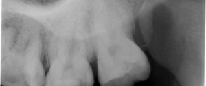
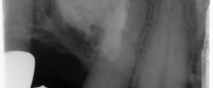
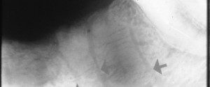
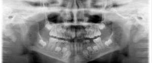
Case of the week: Talon cusp with clinical photo

This week I have a case of a large talon cusp on a maxillary central incisor along with a clinical photo.  A talon cusp is a hyperplasia of the cingulum of a tooth and typically classified as a variant of normal anatomy.  In the anterior, it will present as a…
1 min read 0

Anatomy Monday: Coronoid Process (mandible)

The coronoid process is the superior aspect of the anterior portion of the ramus of the mandible.  It is visible on intraoral radiographs but not mandibular periapical radiographs instead it is seen on maxillary molar periapical radiographs.  This typically due to the posterior placement of the image receptor and holder…
1 min read 0

Case of the week: External resorption of an impacted tooth

This week is a neat case of an impacted maxillary canine with external resorption occurring on the crown.  When looking at these radiographs note that there is no longer an identifiable follicle around the crown of the tooth.  Also note that the areas where there is tooth loss of the…
1 min read 0

Anatomy Monday: Vascular/Nutrient Canals (mandible)

This Monday I am showing vascular canals or nutrient canals as they are sometimes referred to. While they are in both the maxilla and mandible, they are more commonly seen in the mandible particularly the anterior. This is due to thin bone in this area. They will appear as radiolucent…
1 min read 0

Case of the week: Supernumerary tooth

Case of the week is back. I know it’s been a little while but this is a fun one.  This week I have a case of a supernumerary tooth in hiding.  Well, it’s sort of in hiding.  First take a look at the pantomograph and see if you can find…