1 min read
0
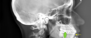
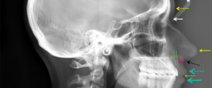
Lateral Cephalometric Skull Anatomy – Part VI
This is the final post and it will cover soft tissue anatomy seen on lateral cephalometric skull radiographs. Glabella (yellow dotted arrow) Nasion (white solid arrow) Pronasale (yellow solid arrow) Subnasale (black dotted arrow) Labrale superius (turquoise dotted arrow) Stomion (green solid arrow) Labrale inferius (turquoise solid arrow) Gnathion (white…