1 min read
0
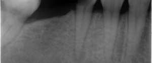
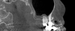
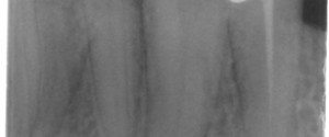
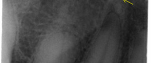
Case of the Week: Oligodontia
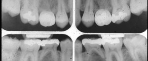
This week I have a case of developmentally missing teeth (oligodontia) along with the last educational video in my library (to date). 🙂 This case shows all four second premolars did not develop with retained primary second molars. And for the last of this years educational videos on oligodontia by…