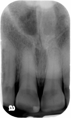
This week I have a case of attrition along with an educational video. First the case; this is an example of attrition on the lingual surfaces of the maxillary incisors. It presents with the crown having an increased radiolucent appearance and a defined horizontal line near the cemento-enamel junction where the attrition stops at.
 Bonus question – what is the radiopaque entity at the superior aspect of the radiograph?
And now for the educational video by Dental Class of 2015 students Todd Herpy and Trino Nuno.
If you have any questions or comments about attrition, please leave them below. Thanks and enjoy!


are they Middle concha?
Could you please tell me where you are looking? Thanks. 🙂
Sorry for the mistaken interpretation. I am looking at the superior aspect only. According to me this radiopacity can be of Maxillary Tori.
You are correct, it is the radiopacity of bone and it is palatal tori. 😀