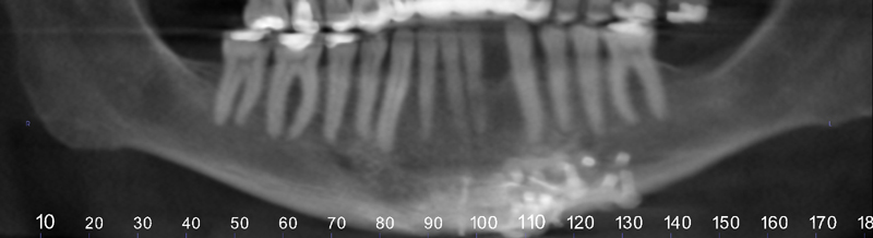
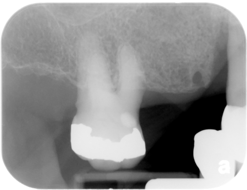
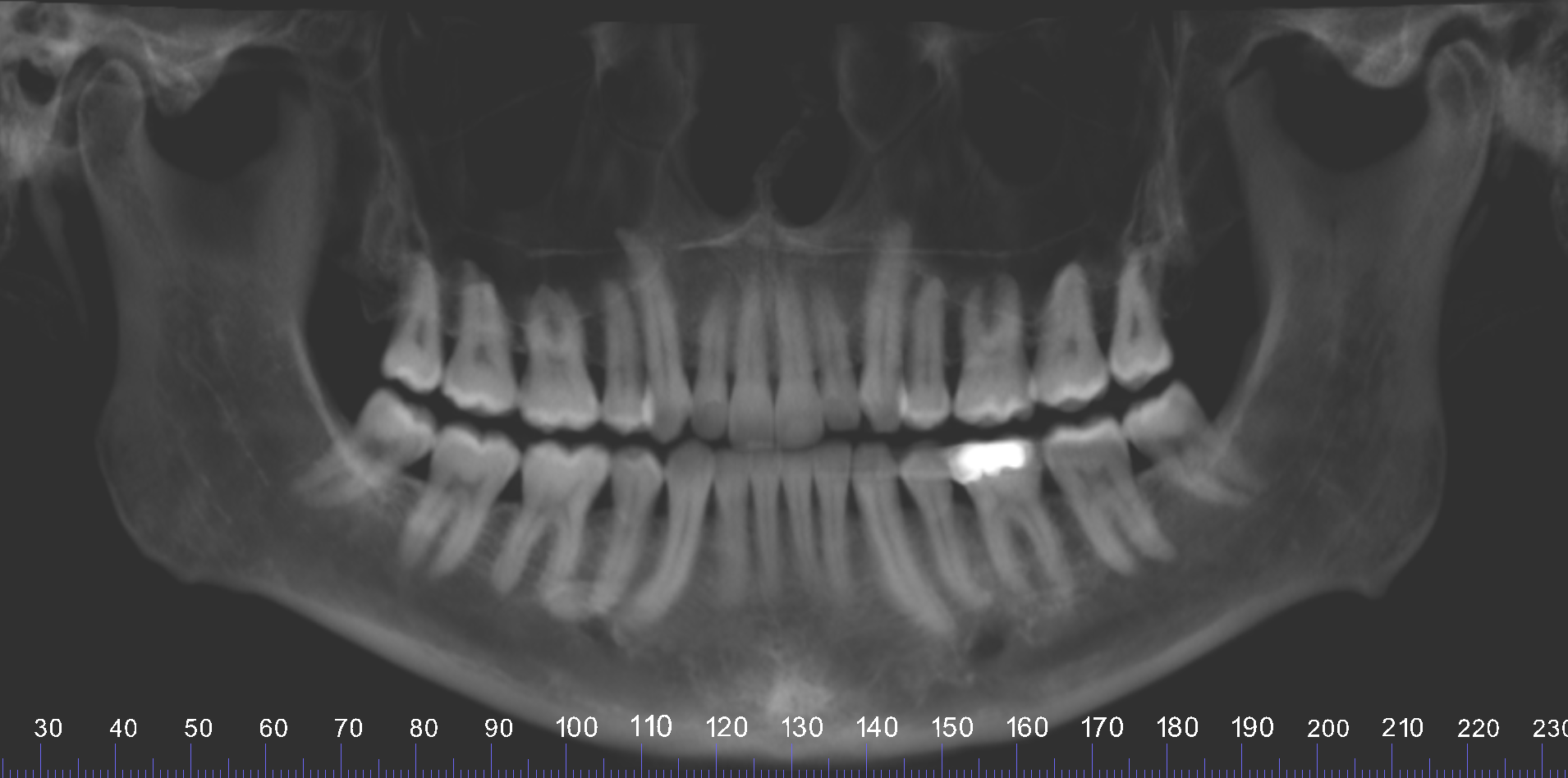
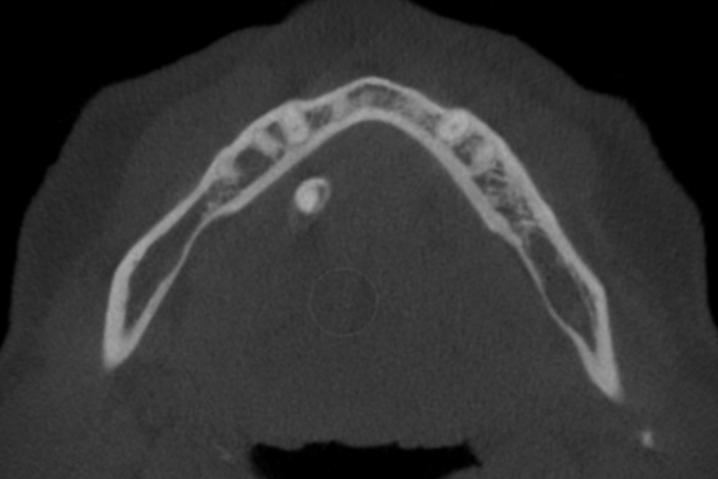
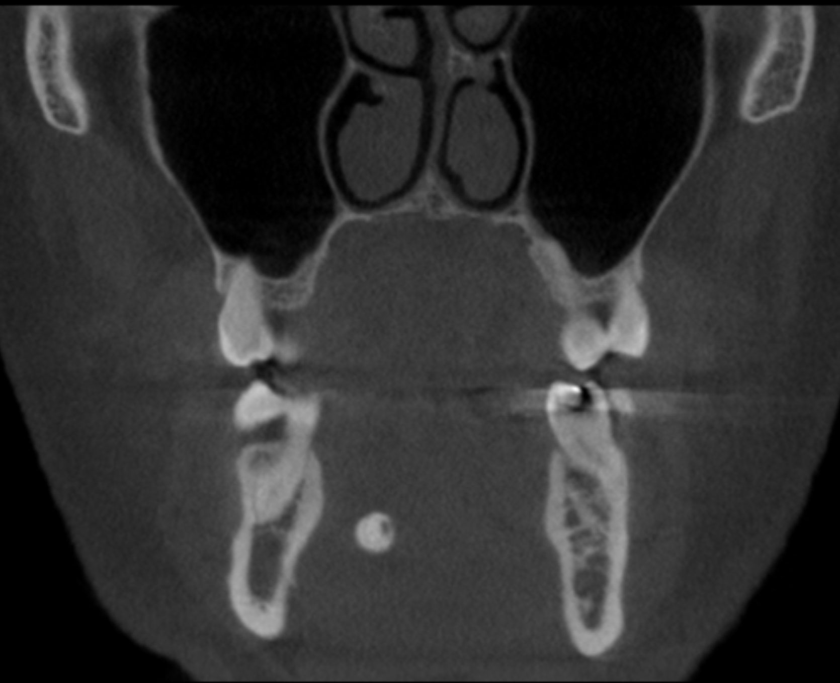
This week I have a case of a sialolith on CBCT (still images and videos) showing the layers of calcifications as it grows. First some information on sialoliths.
QUICK DEFINITION: Sialoliths are calcifications within the ducts of the salivary glands. Most commonly associated with the submandibular salivary gland in the submandibular (Wharton) duct. Patients may be asymptomatic or have swelling especially during eating.
RADIOGRAPHIC DESCRIPTION (LESION) – CBCT:
- Location = floor of the mouth (best visualized on axial and coronal views).
- Edge = well-defined.
- Shape = round/ovoid.
- Internal aspect = radiopaque, may have layers (laminated appearance – looks like the inside of an onion).
- Other = N/A.
- Number = typically single.
Thanks and enjoy!