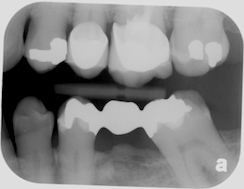
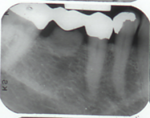
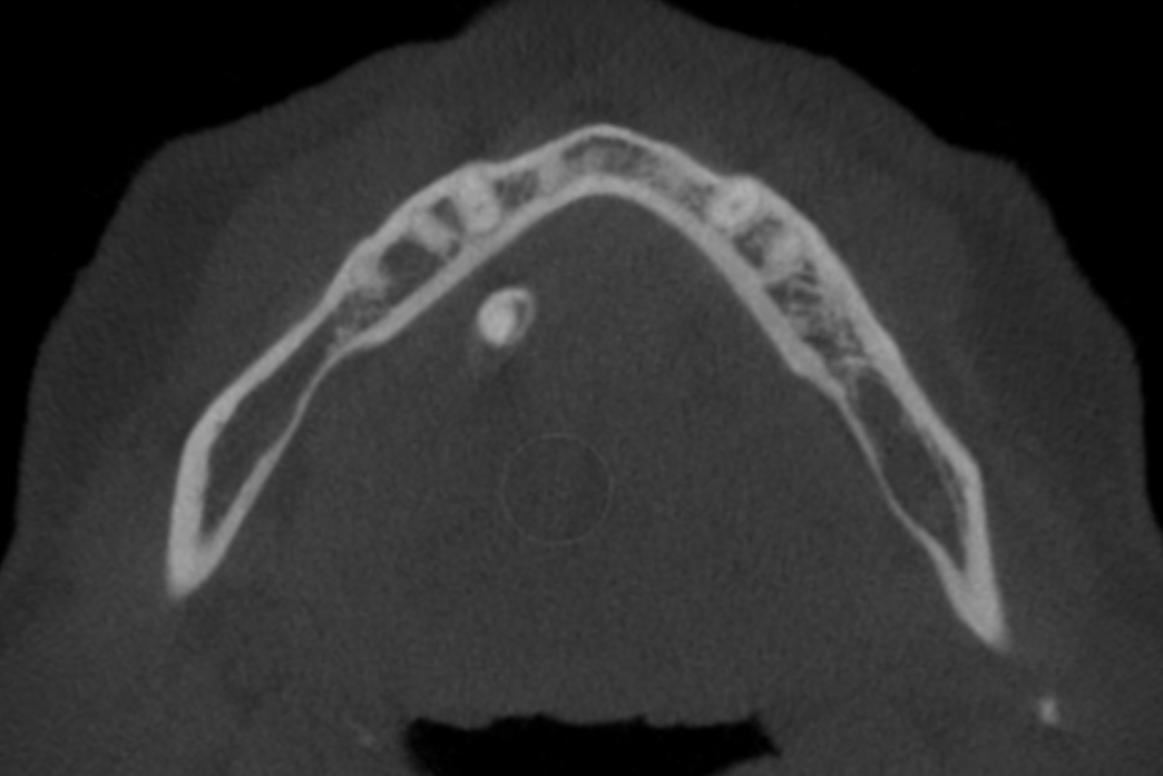
This week I’m sharing examples of excess bone growth in response to a pontic – subpontic osseous hyperplasia or subpontic hyperostosis. There have been some theories as to etiologies including a low grade inflammatory process causing the body to create more bone that can reach up to the pontic in some cases. A few reported cases show that if the pontic is removed the bone will slowly atrophy back to normal limits and in one case when a new pontic was placed the excess bone growth came back. I find it really interesting. 😀
Here are some examples (sorry for the small images).


If you have any questions or comments, please leave them below. Thanks and enjoy!

Wow. I’ve never seen this before.
Thanks for posting it! 🙂
You’re welcome. 🙂
Any theory as to why it happens rather than the opposite?
Some have hypothesized that it may be due a low grade inflammatory response and that in those certain individuals the body responds by creating more bone instead of moving away from the inflammation. It is an interesting response whatever the cause may be.
One of my mentors Dr Walter Daniels published a paper on this topic. Very interesting.
It is a fun, interesting find for sure. 🙂