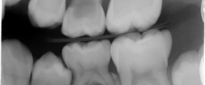
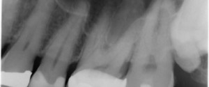
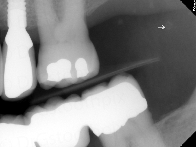
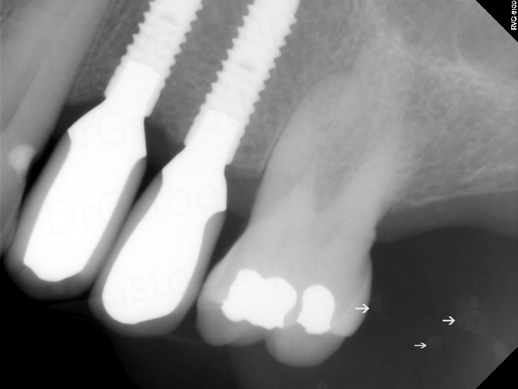
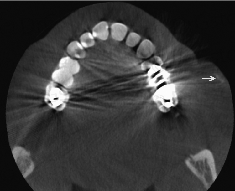
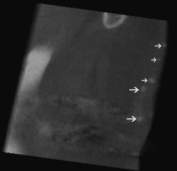
This week I have a fun case of osteoma cutis on both 2D images and CBCT images. Osteoma cutis is an ossification within the dermis. It presents on 2D radiographs as punctate, round/ovoid, radiopaque entities. Most of the cases I come across it occurs in multiples. First is the 2D radiographs followed by the CBCT views.
If you have any questions or comments, please leave them below. Thanks and enjoy!