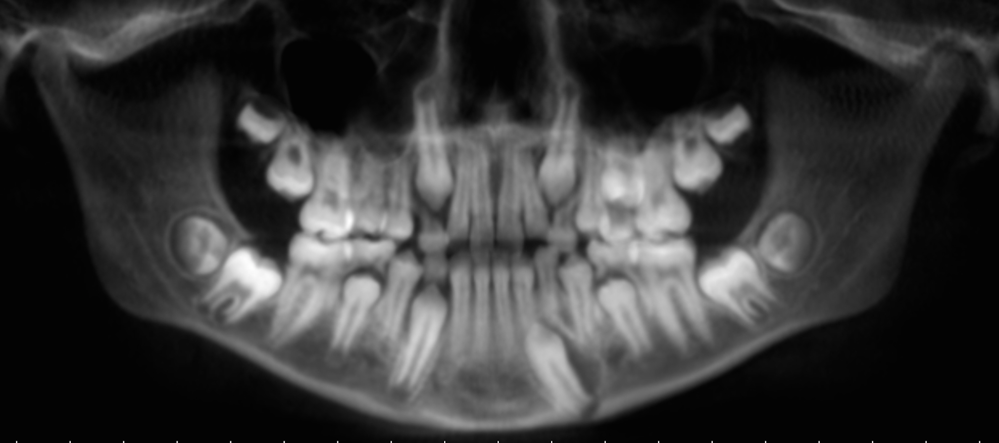
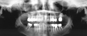
This week I have an interesting case of a lateral dentigerous cyst to share. A lateral dentigerous cyst is a dentigerous cyst that is coming off the lateral aspect of a tooth. As with a typical dentigerous cyst, you will be looking for an enlargement of the dental follicle surrounding an impacted tooth.
This case is on the developing permanent mandibular left canine (#22). Check out the images.
If you have any questions or comments, please leave them below. Thanks and enjoy!
Did the patient have any symptom? Or was an incidental finding?
Incidental finding.
Very nice indeed.
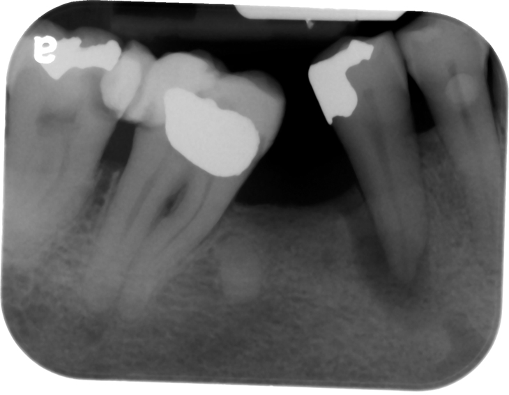
Was a regular OPG done earlier for a screening?
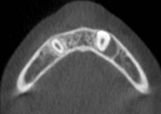
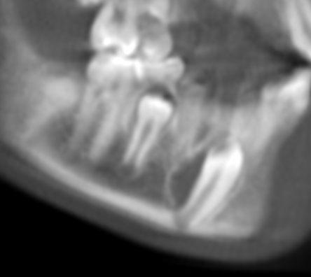
Why was the CBCT done for the patient?
the reason I am asking is – this shows the usefulness of a 3D image in contrat to a 2D image where such things may not be visible at all
I do not know the history but do know the scan was taken to evaluate the canine area. This makes it sound like it was found initially on 2D imaging.