1 min read 2

Anatomy Monday: Pineal Gland Calcification

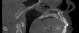
This week I’m onto a calcification encountered in the soft tissue of the brain; the pineal gland.  It appears as a well-defined radiopaque entity in the mid-line of the brain superior to the spinal column. Another entity from my book (Interpretation Basics of Cone Beam Computed Tomography). If you have…
1 min read 5

Case of the Week: Pulp Stones

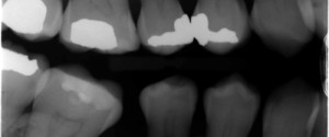
This is an interesting case of multiple teeth with pulp stones. Pulp stones present as well-defined radiopaque entities in the pulp chamber and/or root canal space.  If they are large enough, the canal will be enlarged around them to accommodate them.  Check out the pulp stone party below. 😀 If…
1 min read 0

Anatomy Monday: Spheno-Occipital Synchondrosis

Onto another fun entity shown in my new book (Interpretation Basics of Computed Tomography); the spheno-occipital synchondrosis. The spheno-occipital synchondrosis is a normal developmental entity seen as a well-defined discontinuity of the sphenoid bone in children and teens.   If you have any questions or comments about the spheno-occipital synchondrosis,…
1 min read 3

Case of the Week: Dens Evaginatus

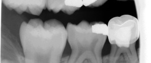
This week is a really cool case of a dens evaginatus erupting onto the plane of occlusion.  Dens evaginatus is an outfolding of the enamel from the occlusal surface of a tooth, which is opposite of dens invaginatus.  On this case, look at the mandibular second premolar which appears to…
1 min read 0

Anatomy Monday: Concha Bullosa

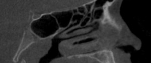
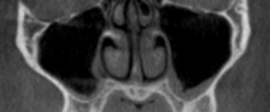
I’ve decided to mix up the Anatomy Monday a little this month celebrating the release of my book – Interpretation Basics of Cone Beam Computed Tomography 😀 by covering entities shown in the book. Today is concha bullosa, an aerated concha most commonly associated with the middle concha. It is…