Being that I’ve been asked quite a few questions on cervical burnout versus caries, I thought it might help to go over the topic a little more in depth. Here are the initial four caries interpretation posts (interproximal caries, occlusal caries, recurrent caries, root caries). Here’s the goods on cervical burnout.
- Presents as a diffuse radiolucent area on the interproximal surfaces of the root apical to the cemento-enamel junction.
- Created by decreased x ray attenuation of the cementum as it is very thin and unable to stop x rays from reaching the image receptor.
- No visible tooth structure missing in this area.
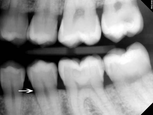
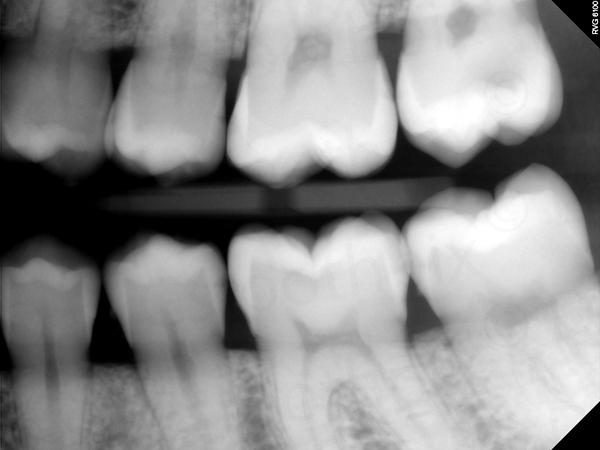
Using some of the previous find the caries examples, see if you can find other areas of cervical burnout.
Please leave any questions or answers in the comments below. Thanks and enjoy!


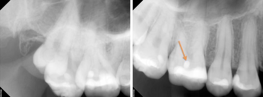
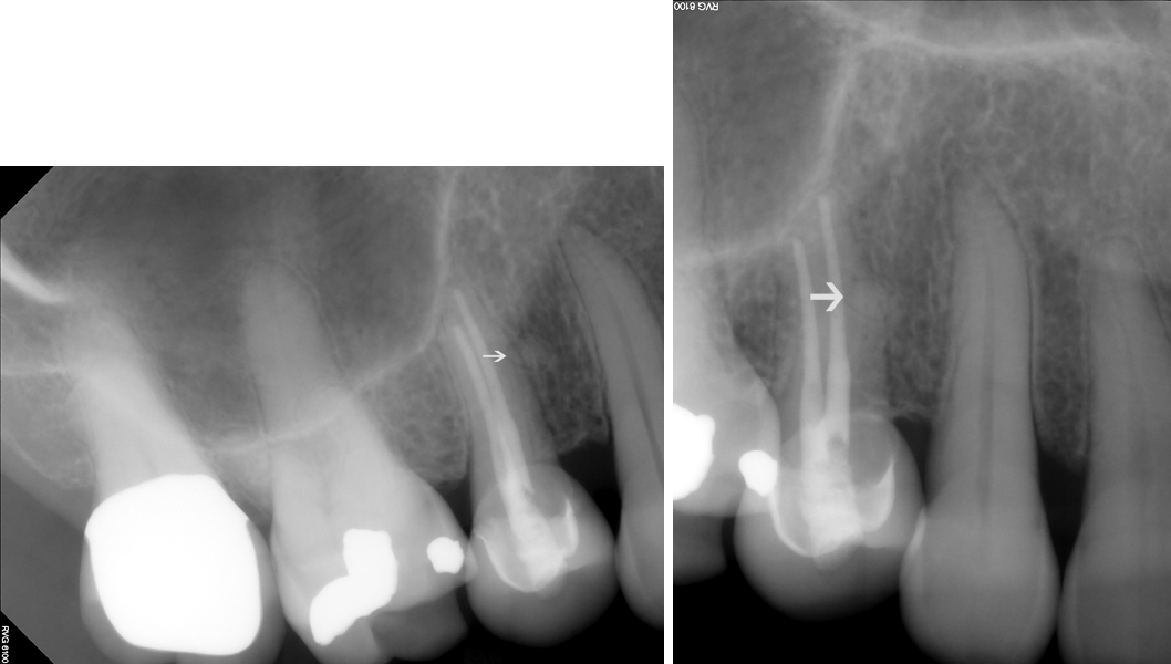
Hi! Will cervical burnout be seen regularly at the same spots of the same teeth, or is it an artefact that depends on film positioning etc.? Is a radiolucency occuring repeatedly in radiographs near the CEJ (eg last row right bitewing, mesial of lower second premolar and for left bitewing, distal of mandibular second premolar) enough evidence to suspect interproximal caries?
Cervical burnout will be seen in the same area (just inferior to CEJ). It can occur on any tooth but is more commonly seen on premolars. When evaluating for caries near the CEJ, look for actual tooth structure missing. If you are using digital you can lighten the image to see if the normal shape of the crown and root exist in this location (i.e. cervical burnout) versus caries. I hope this helps. 🙂