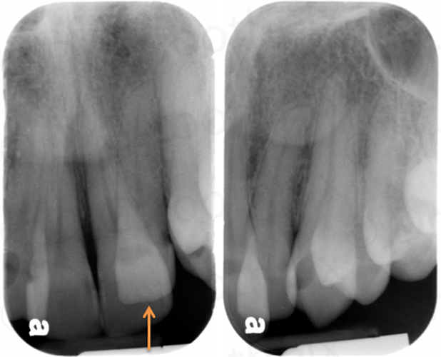
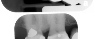
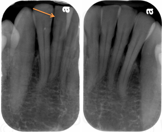
And now the answers for the October 2014 Find the Caries.
- maxillary right second premolar (#4) – distal
- mandibular left second molar (#18) – mesial
- mandibular right second premolar (#29) – distal
There are a few other spots that appear questionable. What other surfaces are catching your eye?
If you have any questions or answers, please leave them below. Thanks and enjoy!


I would not treat any of these teeth based on these radiographic images. None of the interproximal decalcifications appear in my view to extend axially past the DEJ. I would monitor with yearly BW radiographs, give home care instructions specifically focused on interproximal hygiene, and prescribe Prevident Fluoride toothpaste. Ed
Ed – Thanks for the clinical insight on these lesions. I agree that they are quite small. I purposely do not go into treatment one these posts but use them as practice for others to quiz themselves on radiographic caries interpretation. Thanks again. 🙂
I think there is enamel caries #30 M , #31 M
Yes, I would agree that the mesial surfaces of both # 30 and # 31 have carious lesions on them.
how about occlusal seconcaries on #45?
I am enjoying your website. Thank you a lot.
I mean on lower second premolar
Thanks and I’m glad you are finding the website useful.
As for the occlusal of the mandibular right second premolar, if you are referring to the well-defined radiolucent area under the existing occlusal restoration that is a void where the material was not adequately packed down.
how about mesial of 3.7?
is it also void on occlusal of 3.6?